Review
. 2019 Feb 7;20(3):718.
doi: 10.3390/ijms20030718.

Interactions between Host Immunity and Skin-Colonizing Staphylococci: No Two Siblings Are Alike

Affiliations
Review

Interactions between Host Immunity and Skin-Colonizing Staphylococci: No Two Siblings Are Alike

Young Joon Park et al. Int J Mol Sci. .

Abstract

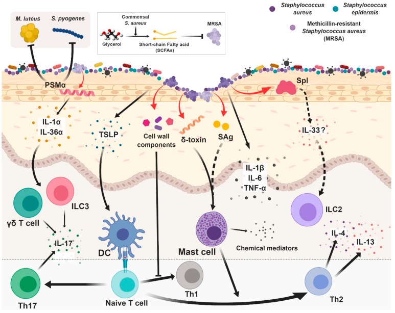
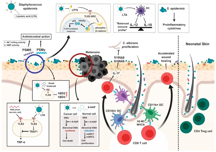
As the outermost layer of the body, the skin harbors innumerable and varied microorganisms. These microorganisms interact with the host, and these interactions contribute to host immunity. One of the most abundant genera of skin commensals is Staphylococcus. Bacteria belonging to this genus are some of the most influential commensals that reside on the skin. For example, colonization by Staphylococcus aureus, a well-known pathogen, increases inflammatory responses within the skin. Conversely, colonization by Staphylococcus epidermis, a coagulase-negative staphylococcal species that are prevalent throughout the skin, can be innocuous or beneficial. Thus, manipulating the abundance of these two bacterial species likely alters the skin microbiome and modulates the cutaneous immune response, with potential implications for various inflammation-associated skin diseases. Importantly, before researchers can begin manipulating the skin microbiome to prevent and treat disease, they must first fully understand how these two species can modulate the cutaneous immune response. In this review, we discuss the nature of the interactions between these two bacterial species and immune cells within the skin, discussing their immunogenicity within the context of skin disorders.

Keywords: Staphylococcus aureus; Staphylococcus epidermis; Staphylococcus spp., T cells; atopic dermatitis; commensals; cutaneous immunity; microbiome.


Conflict of interest statement

The authors declare no conflict of interest.

Figures

Figure 1
Figure 1
Interaction between Staphylococcus aureus and skin immune system. The skin immune system comprises a complex network of cells. Because keratinocytes serve as sensors, phenol-soluble modulin α (PSMα) are capable of inducing epidermal keratinocytes to produce IL-1 and IL-36, even without epidermal disruption. T cells and innate lymphoid cells (ILC3) containing the IL-36 receptor recognize IL-13 and secrete IL-17, leading to inflammation. Another peptide belonging to the PSM family, δ-toxin, promotes Th2 responses by activating mast cells. It is likely that staphylococcal superantigens (SAgs) are also capable of mast cell activation. Thymic stromal lymphopoietin (TSLP) can drive Th2 skewing via dendritic cells (DCs). Cell wall components from S. aureus inhibit Th1 responses. S. aureus also possesses probiotic properties. S. aureus shows antimicrobial activity against other bacteria, such as M. luteus and S. pyogenes. Commensal S. aureus inhibits methicillin-resistant S. aureus (MRSA) using short-chain fatty acids (SCFAs) fermented from glycerol. Red arrows indicate bacteria-originated materials. T-bar indicates inhibitory activity.
Figure 2
Figure 2
Interaction between Staphylococcus epidermis and skin immune system. When skin is injured, lipoteichoic acid (LTA) from the skin commensal S. epidermidis inhibits inflammatory cytokine production via inhibition of TLR3 signaling by TRAF1 activated through TLR2. S. epidermidis also produces small molecules that increase anti-microbacterial peptides through TLR2, which in turn inhibits pathogen growth. Lipopeptide 78 activates TLR2-SRC signaling in keratinocytes to induce β-catenin phosphorylation. Dissociation between p65 and PPARγ due to phospho-β-catenin binding reduces TLR3-mediated inflammation. Phenol-soluble modulin γ (PSMγ) and PSMδ from S. epidermidis also have anti-microbacterial activity. 6-HAP, a nucleobase analogue capable of selective tumor growth suppression was recently found to be produced by S. epidermidis. Cancer cells did not have sufficient mARC1 and mARC2 to reduce 6-HAP, leading to cell death. CD103+ dendritic cells (DCs) and CD11b+ DCs cooperate in tuning S. epidermidis-specific CD8+ T cells to protect from pathogen infection via upregulation of alarmins (S100A8 and S100A9). The commensal-specific CD8+ T cell response is induced by non-classical MHC class I molecules (H2-M3 MHCIb), and is immunoregulatory and tissue-repairing in nature. Dendritic cells (DCs) exposed to LTA preparations from S. epidermidis (epi-LTA) show an IL-10-balanced immune profile, with the ratio of IL-12p70 to IL-10 being close to 1; likewise, naïve CD4+ T cells primed by epi-LTA produce lower amounts of IFN-γ and IL-17. In neonatal skin, S. epidermidis is critical for the accumulation of CD4+ regulatory T cells, establishing tolerance to skin commensals. Even in low amounts, the inflammatory cytokines induced by S. epidermidis can lead to various consequences, such as sepsis, bronchopulmonary dysplasia, white matter injury, necrotizing enterocolitis, and retinopathy of prematurity.

Similar articles

Cited by

References

    1. Hand T.W. The Role of the Microbiota in Shaping Infectious Immunity. Trends Immunol. 2016;37:647–658. doi: 10.1016/j.it.2016.08.007. - DOI - PMC - PubMed
    1. Lamers R.P., Muthukrishnan G., Castoe T.A., Tafur S., Cole A.M., Parkinson C.L. Phylogenetic relationships among Staphylococcus species and refinement of cluster groups based on multilocus data. BMC Evol. Biol. 2012;12:171. doi: 10.1186/1471-2148-12-171. - DOI - PMC - PubMed
    1. Krismer B., Weidenmaier C., Zipperer A., Peschel A. The commensal lifestyle of Staphylococcus aureus and its interactions with the nasal microbiota. Nat. Rev. Microbiol. 2017;15:675–687. doi: 10.1038/nrmicro.2017.104. - DOI - PubMed
    1. Totte J.E., van der Feltz W.T., Hennekam M., van Belkum A., van Zuuren E.J., Pasmans S.G. Prevalence and odds of Staphylococcus aureus carriage in atopic dermatitis: A systematic review and meta-analysis. Br. J. Dermatol. 2016;175:687–695. doi: 10.1111/bjd.14566. - DOI - PubMed
    1. Higaki S., Morohashi M., Yamagishi T., Hasegawa Y. Comparative study of staphylococci from the skin of atopic dermatitis patients and from healthy subjects. Int. J. Dermatol. 1999;38:265–269. doi: 10.1046/j.1365-4362.1999.00686.x. - DOI - PubMed