. 2019 Feb 8;24(1):10.
doi: 10.1186/s40001-019-0366-9.

Human umbilical cord mesenchymal stem cells implantation accelerates cutaneous wound healing in diabetic rats via the Wnt signaling pathway

Affiliations

Human umbilical cord mesenchymal stem cells implantation accelerates cutaneous wound healing in diabetic rats via the Wnt signaling pathway

Yanfu Han et al. Eur J Med Res. .

Abstract

Objective: Difficulty in wound healing is one common complication of diabetes mellitus. The study explored whether the therapeutic effect of human umbilical cord mesenchymal stem cells (hUCMSCs) on diabetic ulcer wound was enhanced by the activation of the Wnt signaling pathway.

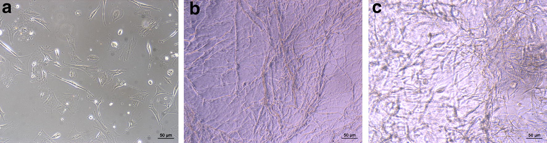
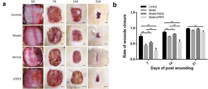
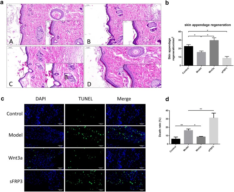
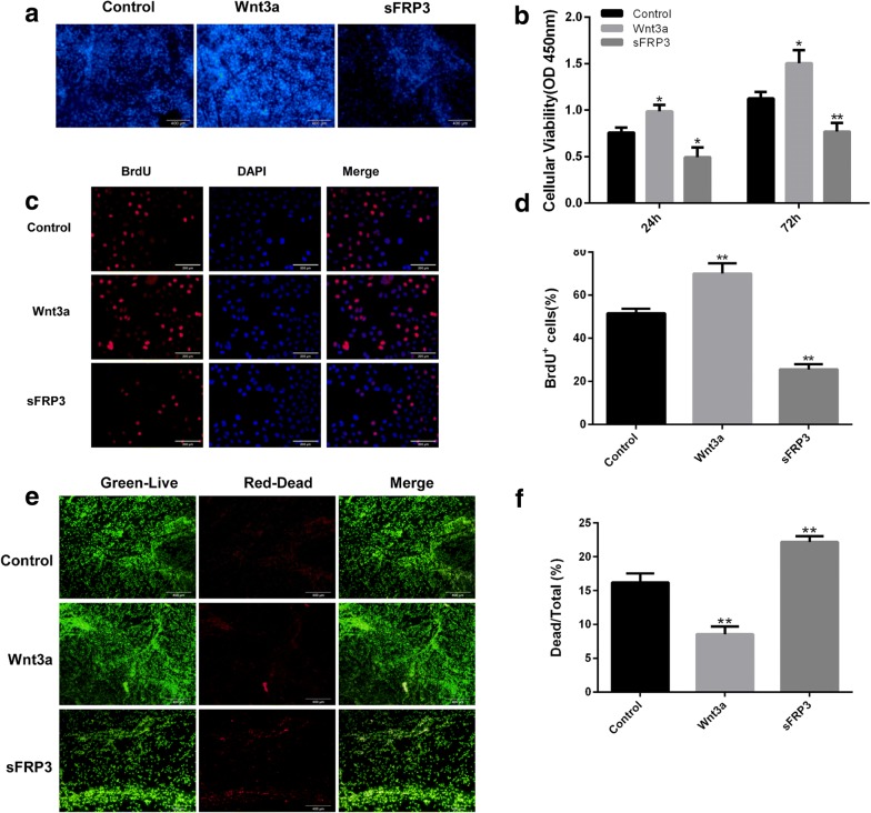
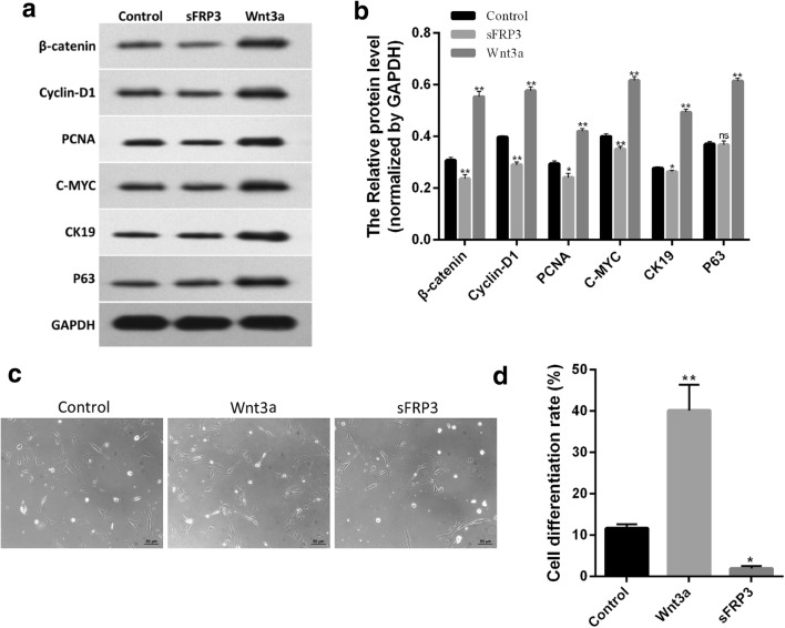
Methods: Rat diabetic model was established by intraperitoneal injection of Streptozotocin (STZ). hUCMSCs were purified and seeded on the collagen-chitosan laser drilling acellular dermal matrix (CCLDADM) scaffold, which was subsequently implanted into the cutaneous wound of normal and diabetic rats, followed by daily injection of Wnt signaling pathway agonist (Wnt3a) or antagonist (sFRP3) at the edge of the scaffold. Wound healing was checked on days 7, 14, and 21, and the fibrous tissue deposition, capillaries, and epidermal regeneration at the wound were examined by hematoxylin-eosin staining. The hUCMSCs-CCLDADM scaffold was cultured in vitro and treated with Wnt3a or sFRP3, followed by evaluation of cell proliferation, cell proliferation rate, survival status, and altered protein levels in the Wnt signaling pathway using BrdU staining, CCK-8 assay, live/dead staining, and Western blotting, respectively.

Results: On days 7 and 14 postoperatively, the speed of wound healing was significantly lower in diabetic rats than that in normal control rats. This phenomenon was significantly improved by the activation of the Wnt signaling pathway that also elevated the fibrous protein deposition and the abundance of capillary in the granulation tissue. Conversely, blockade of Wnt signaling slowed the healing of skin wound in diabetic rats. The activation of Wnt signaling pathway promoted the proliferation and differentiation and decreased the apoptosis of hUCMSCs, thereby elevating the number of living hUCMSCs on the CCLDADM scaffold, while the suppression exerted a contrary effect.

Conclusion: The activation of the Wnt signaling pathway promotes the healing of diabetic skin wound by the regulation of proliferation and differentiation of hUCMSCs on the CCLDADM scaffold.

Keywords: Diabetes mellitus; Mesenchymal stem cell therapy; Tissue engineering skin; Wnt signaling pathway; Wound healing.


Figures

Fig. 1
Fig. 1
hUCMSCs morphology and growth characteristics on the CCLDADM scaffold. a hUCMSCs morphology under optical microscope (scale: 50 μm). b CCLDADM scaffold shape (scale: 50 μm). c Morphology of hUCMSCs after seeding on the CCLDADM scaffold (scale: 50 μm)
Fig. 2
Fig. 2
Activation of the Wnt signaling pathway improved the therapeutic effect of hUCMSCs-loaded scaffold for diabetic wounds. a Wound healing in different intervention groups on days 7, 14, and 28 postoperative (scale: 0.25 cm). b Statistical analysis of results in c (n = 6); *P < 0.05, **P < 0.01
Fig. 3
Fig. 3
Activation of the Wnt signaling pathway promoted the generation of granulation tissues. a HE staining demonstrated the collagen fiber deposition and changes in capillaries in the granulation tissues (scale: 100 μm). b Statistical analysis of a results. c TUNEL staining detected cell apoptosis at the wound (scale: 50 μm). d Statistical analysis of c results (n = 3); *P < 0.05, **P < 0.01
Fig. 4
Fig. 4
Activation of the Wnt signaling pathway promoted hUCMSCs survival on the CCLDADM scaffold. a hUCMSCs were seeded on the CCLDADM scaffold and treated with Wnt3a or sFRP3 for 72 h, followed by DAPI staining to estimate the total number of cells on the scaffold (scale: 200 μm). b CCK-8 staining detected the total number of cells on the CCLDADM scaffold at 24 or 72 h post-drug intervention (n = 3). c hUCMSCs were seeded in a 6-well plate and treated for 72 h with Wnt3a or sFRP3, and the ratio of BrdU-positive cells was detected to reflect the rate of proliferation (scale: 100 μm). d Statistical analysis of BrdU-positive ratio from c (n = 3). e Live/dead cell staining evaluated the ratio of dead cells on the CCLDADM scaffold (scale: 100 μm). f Statistical analysis of results from e (n = 3); *P < 0.05, **P < 0.01 vs. control
Fig. 5
Fig. 5
Activation of the Wnt signaling pathway enhanced hUCMSCs differentiation. a Western blotting was applied to detect the changes in the levels of β-catenin, c-Myc, p63, CK19, and PCNA proteins. b Statistical analysis of a results. c The differentiation degree of hUCMSCs was evaluated by cell morphology under optical microscope (scale: 100 μm). d Statistical analysis on the ratio of cells with round or small polygonal morphology displayed in c; *P < 0.05, **P < 0.01 vs. control (n = 3)

References

    1. Jiang Y, Ran X, Jia L, Yang C, Wang P, Ma J, et al. Epidemiology of type 2 diabetic foot problems and predictive factors for amputation in China. Int J Low Extrem Wounds. 2015;14:19–27. doi: 10.1177/1534734614564867. - DOI - PubMed
    1. Hu C, Jia W. Diabetes in China: epidemiology and genetic risk factors and their clinical utility in personalized medication. Diabetes. 2018;67:3–11. doi: 10.2337/dbi17-0013. - DOI - PubMed
    1. Wang Y, Chen X, Cao W, Shi Y. Plasticity of mesenchymal stem cells in immunomodulation: pathological and therapeutic implications. Nat Immunol. 2014;15:1009–1016. doi: 10.1038/ni.3002. - DOI - PubMed
    1. Ma S, Xie N, Li W, Yuan B, Shi Y, Wang Y. Immunobiology of mesenchymal stem cells. Cell Death Differ. 2014;21:216–225. doi: 10.1038/cdd.2013.158. - DOI - PMC - PubMed
    1. Ding DC, Chang YH, Shyu WC, Lin SZ. Human umbilical cord mesenchymal stem cells: a new era for stem cell therapy. Cell Transplant. 2015;24:339–347. doi: 10.3727/096368915X686841. - DOI - PubMed

LinkOut - more resources