Review
. 2019 Apr;25(4):328-340.
doi: 10.1016/j.molmed.2019.01.002. Epub 2019 Feb 5.

Macrophages in Heart Failure with Reduced versus Preserved Ejection Fraction

Affiliations
Review

Macrophages in Heart Failure with Reduced versus Preserved Ejection Fraction

Matthew DeBerge et al. Trends Mol Med. 2019 Apr.

Abstract

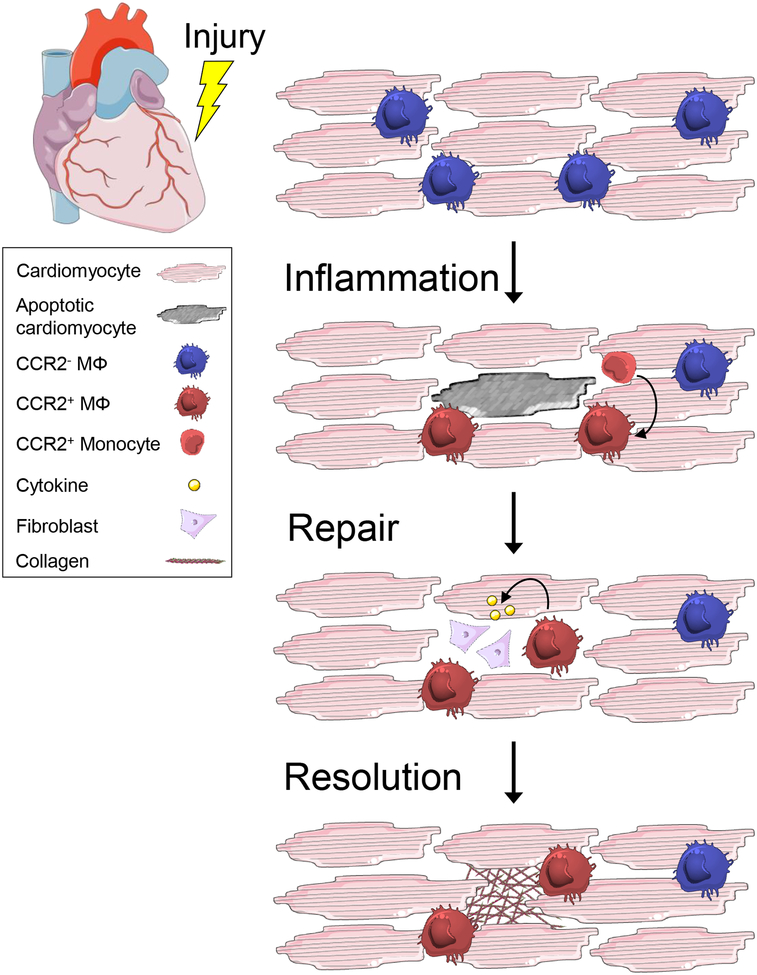
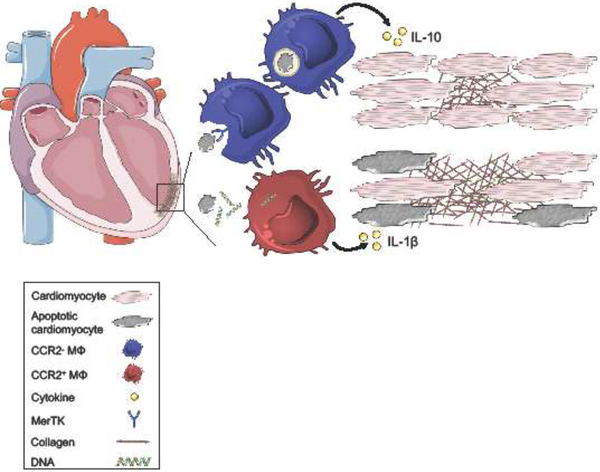
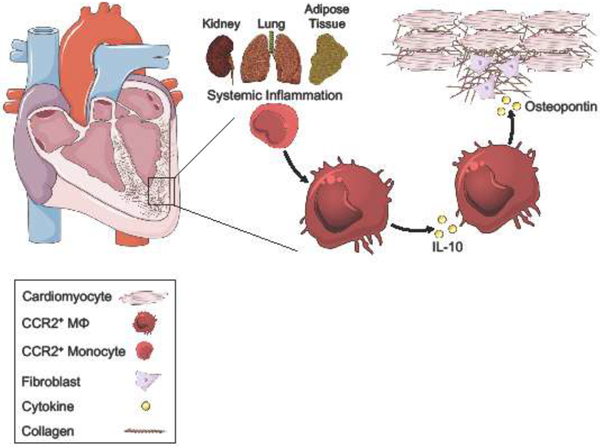
There is a growing number of individuals living with heart failure (HF) with reduced ejection fraction (HFrEF) or preserved ejection fraction (HFpEF). Long-term prognosis remains poor in both cases, especially in HFpEF, which is rising in incidence and lacks effective therapeutics. In both HFrEF and HFpEF, there is evidence that elevated inflammatory biomarkers, implicating innate immune cells such as macrophages, are associated with worsened clinical outcomes. Macrophage subsets are active in both inflammatory and reparative processes, yet our understanding of the causative roles for these cells in HF development and progression is incomplete. Here, we discuss recent findings interrogating the role of macrophages in inflammation and its resolution in the context of HF, with a specific focus on HFrEF versus HFpEF.

Keywords: heart failure; inflammation; macrophage; preserved ejection fraction; reduced ejection fraction.


Figures

Figure 1.
Figure 1.. Inflammation Resolution is an active process.
Tissue repair is characterized by 3 phases: inflammation, repair, and resolution. Inflammation can be initiated both locally or systemically and functions to eliminate the source of injury and remove damaged tissue. In the heart, it is characterized by local cell death, loss of CCR2 resident macrophages, and replacement by recruited CCR2+ monocyte-derived macrophages. Repair is initiated after engulfment and clearance of apoptotic cardiomyocytes, stimulating the release of cytokines, including IL-10 and TGF-β, which activate fibroblasts to replace and remodel the tissue. Resolution is achieved following elimination of the injurious source and clearance of dead cells and debris. It is characterized by apoptosis and egress of the reparative machinery and restoration of homeostasis. In both HFrEF and HFpEF, the failure to resolve inflammation is correlated with pump dysfunction and ultimately, end-stage HF.
Figure 2.
Figure 2.. The balance of macrophage inflammatory and reparative responses sets the clinical trajectory in HFrEF.
Following myocardial infarction, cardiomyocyte death primarily occurs through apoptosis and necrosis. Macrophages express surface receptors, including MER proto-oncogene Tyrosine Kinase (MerTK), that recognize and mediate phagocytosis of apoptotic cells. Engulfment and metabolism of dying cells stimulates macrophage production of anti-inflammatory cytokines, including IL-10, which preserves neighboring tissue and cardiac function. Dying cardiomyocytes also secrete damage-associated molecular patterns (DAMP), including double-stranded DNA. Macrophage recognition of DAMPs promotes secretion of pro-inflammatory cytokines, including IL-1β leading to collateral tissue damage, adverse ventricular remodeling, and systolic dysfunction.
Figure 3.
Figure 3.. Systemic inflammation perpetuates macrophage repair processes in HFpEF.
The risk of HFpEF increases with age and is associated with an increase in the prevalence of extracardiac comorbidities and risk factors, including chronic kidney disease (kidney), chronic obstructive pulmonary disease (lung), and obesity (adipose tissue). Advanced age and multiple comorbidities in HFpEF patients contribute to a heightened level of systemic inflammation, which promotes myocardial infiltration of CCR2+ monocytes and differentiation into cardiac macrophages. Expression of IL-10 by macrophages leads to autocrine induction of osteopontin, a cytokine associated with cardiac fibrosis. Osteopontin activates cardiac fibroblasts, promoting excess collagen deposition and interstitial fibrosis. These pathologic processes impair cardiomyocyte relaxation and contribute to diastolic dysfunction.

References

    1. Benjamin EJ et al. (2018) Heart Disease and Stroke Statistics-2018 Update: A Report From the American Heart Association. Circulation 137 (12), e67–e492. - PubMed
    1. Heidenreich PA et al. (2013) Forecasting the impact of heart failure in the United States: a policy statement from the American Heart Association. Circ Heart Fail 6 (3), 606–19. - PMC - PubMed
    1. Lee DS et al. (2009) Relation of disease pathogenesis and risk factors to heart failure with preserved or reduced ejection fraction: insights from the framingham heart study of the national heart, lung, and blood institute. Circulation 119 (24), 3070–7. - PMC - PubMed
    1. Chiong M et al. (2011) Cardiomyocyte death: mechanisms and translational implications. Cell Death Dis 2, e244. - PMC - PubMed
    1. Yancy CW et al. (2017) 2017 ACC/AHA/HFSA Focused Update of the 2013 ACCF/AHA Guideline for the Management of Heart Failure: A Report of the American College of Cardiology/American Heart Association Task Force on Clinical Practice Guidelines and the Heart Failure Society of America. J Card Fail 23 (8), 628–651. - PubMed