Review
. 2019 Nov-Dec;23(6):459-466.
doi: 10.1016/j.bjpt.2019.01.008. Epub 2019 Feb 2.

Kinesiologic considerations for targeting activation of scapulothoracic muscles - part 1: serratus anterior

Affiliations
Review

Kinesiologic considerations for targeting activation of scapulothoracic muscles - part 1: serratus anterior

Donald A Neumann et al. Braz J Phys Ther. 2019 Nov-Dec.

Abstract

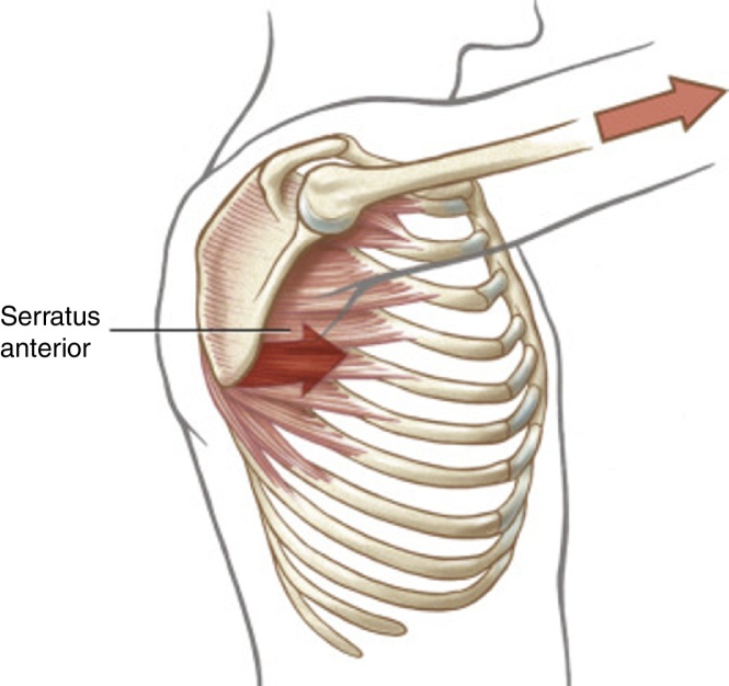
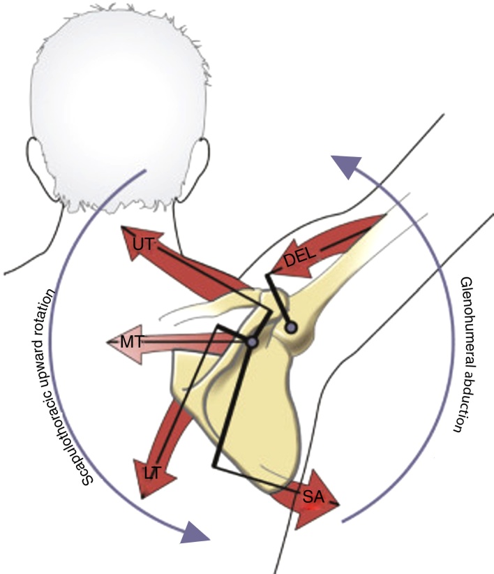
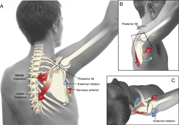
Background: The serratus anterior (SA) is capable of a wide range of actions across the scapulothoracic joint. Furthermore, the lack of control, strength, or activation of this important muscle is believed to be associated with several painful conditions involving the shoulder complex. Studies and clinical intuition have therefore identified several exercises that selectively target the activation of the SA.

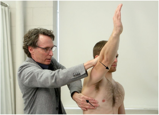
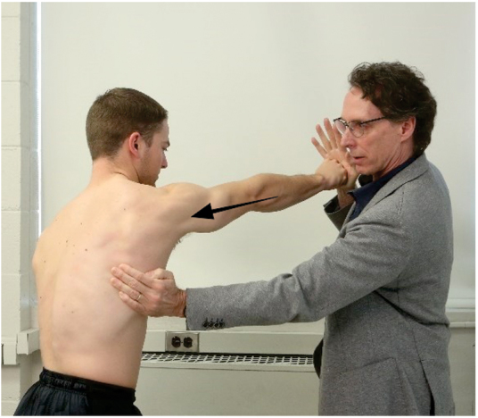
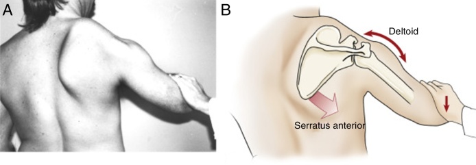
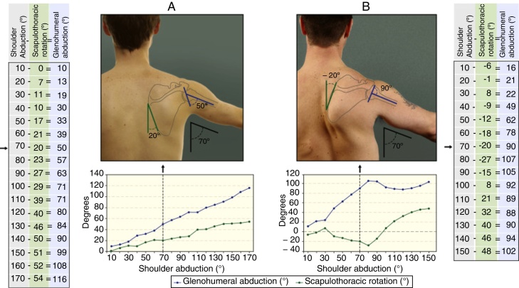
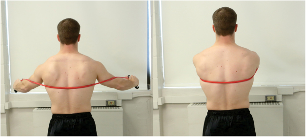
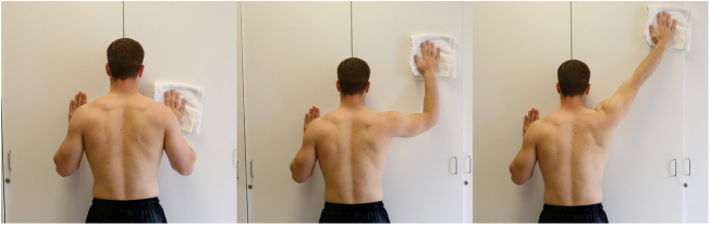
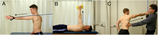
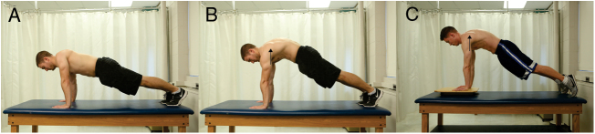
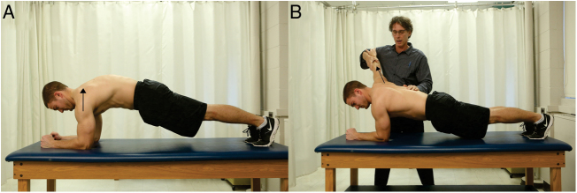
Methods: This paper reviews the anatomy, innervation, testing, and complex actions of the SA. In addition, this paper describes the classic signs and symptoms of weakness or reduced activation of the SA. Several exercises are described and illustrated that purportedly target the activation of the SA, with the intention of optimizing muscular control and encouraging pain free shoulder motion.

Conclusions: This review provides the theoretical background and literature-based evidence that can help explain the SA's complex pathokinesiology, as well as guide the clinician to further develop exercises that likely challenge the muscle. This paper is written along with a companion paper entitled: Kinesiologic considerations for targeting activation of scapulothoracic muscles: part 2: trapezius. Both papers prepare the reader to expand their pallet of exercises that target and challenge these two dominant muscles, with a goal of improving function of the shoulder for several painful conditions caused by their reduced or altered activation pattern.

Keywords: Physical therapy; Scapular dyskinesis; Scapulothoracic joint; Serratus anterior exercises; Shoulder rehabilitation.


Figures

Figure 1
Figure 1
The serratus anterior, showing muscle attachments and overall line of force.
Figure 2
Figure 2
Posterior view of a healthy shoulder during shoulder abduction. The model shows the primary muscle interaction between the scapulothoracic upward rotators and the glenohumeral abductor muscles. The primary force-couple is between the serratus anterior and trapezius. Note two axes of rotation: the scapular axis, located near the acromion, and the glenohumeral joint axis, located at the humeral head. Internal moment arms for all muscles are shown as dark thicker lines. DEL, middle deltoid; LT, lower trapezius; MT, middle trapezius; SA, serratus anterior; UT, upper trapezius.
Figure 3
Figure 3
(A) Theoretical mechanism of how the serratus anterior (SA) and middle trapezius (MT) and lower trapezius (LT) muscles can control the posterior tilt and external rotation of the upwardly rotating scapula during scapular plane abduction. (B) The SA and LT act in a force-couple to posteriorly tilt the scapula relative to the axis of rotation at the acromioclavicular joint (indicated by the green circle). (C) The SA and MT act in a force-couple to externally rotate the scapula relative to the axis of rotation at the acromioclavicular joint (indicated by the blue circle). Each muscle's moment arm is indicated as a dark black line, originating at the axis of rotation of the acromioclavicular joint.
Figure 4
Figure 4
Muscle test for the serratus anterior (shoulder abduction test). The examiner simultaneously resists maximal-effort scapular plane abduction and upward rotation of the scapula.
Figure 5
Figure 5
Muscle test for the serratus anterior (scapular protraction test). The examiner resists maximal-effort protraction of the scapula and the entire upper extremity.
Figure 6
Figure 6
The pathomechanics of the winging right scapula after paralysis of the right serratus anterior caused by an injury of the long thoracic nerve. (A) The scapula is its downwardly rotated position, anteriorly tilted, and internally rotated. (B) Kinesiologic analysis of the extreme downward rotated position. Without an adequate upward rotation force from the serratus anterior (fading arrow), the scapula is not properly stabilized and cannot resist the pull of the deltoid. Subsequently the force of the deltoid (bidirectional arrow) causes the combined actions of downward rotation of the scapula and partial elevation (abduction) of the humerus.
Figure 7
Figure 7
Goniometric measurements used to estimate glenohumeral joint abduction (blue) as the difference between shoulder abduction (black; plotted on the horizontal axis of the graphs) and the scapulothoracic rotation position (green). A healthy male (A) and a male with scapular dyskinesis (B) are each shown holding their shoulder abducted to 70 degrees. Upward rotation of the scapula is indicated by positive angles; downward rotation by negative angles.
Figure 8
Figure 8
Dynamic hug exercise. Using elastic material as resistance, the subject performs bilateral, maximum scapular protraction.
Figure 9
Figure 9
Towel-wall slide. The standing subject slides the towel against a wall, starting in a neutral shoulder position, and ending in combined position of maximal scapular plane abduction and scapular protraction.
Figure 10
Figure 10
Serratus punches: Exercise performed standing (A) or supine (B) against elastic or weight of dumbbell, or against manual resistance (C) by the examiner.
Figure 11
Figure 11
Push-up plus performed on hands and toes: scapulothoracic protraction (thorax moving on fixed scapulae). Initial position (A) and final position (B). Performed on an unstable surface (C).
Figure 12
Figure 12
Push-up plus on elbows and toes: (A) bilateral, scapulothoracic protraction (thorax moving on fixed scapulae). (B) Left side shows subject holding relative scapulothoracic protraction; right side shows examiner resisting scapulothoracic protraction (arm and scapula protracting relative to fixed thorax).

References

    1. Camargo P.R., Neumann D.A. Kinesiologic considerations for targeting activation of scapulothoracic muscles - Part 2: trapezius. Braz J Phys Ther. 2019;23(6):467–475. - PMC - PubMed
    1. Neumann D.A. 3rd ed. Elsevier; St Louis: 2017. Kinesiology of the Musculoskeletal System: Foundations for Rehabilitation.
    1. Standring S. 41st ed. Elsevier; St Louis: 2016. Gray's Anatomy: The Anatomical Basis of Clinical Practice.
    1. Berthold J.B., Burg T.M., Nussbaum R.P. Long thoracic nerve injury caused by overhead weight lifting leading to scapular dyskinesis and medial scapular winging. J Am Osteopath Assoc. 2017;117(2):133–137. - PubMed
    1. Friedenberg S.M., Zimprich T., Harper C.M. The natural history of long thoracic and spinal accessory neuropathies. Muscle Nerve. 2002;25:535–539. - PubMed