. 2019 Feb 19;50(2):477-492.e8.
doi: 10.1016/j.immuni.2019.01.006. Epub 2019 Feb 5.

Clonal Deletion of Tumor-Specific T Cells by Interferon-γ Confers Therapeutic Resistance to Combination Immune Checkpoint Blockade

Affiliations

Clonal Deletion of Tumor-Specific T Cells by Interferon-γ Confers Therapeutic Resistance to Combination Immune Checkpoint Blockade

Chien-Chun Steven Pai et al. Immunity. .

Abstract

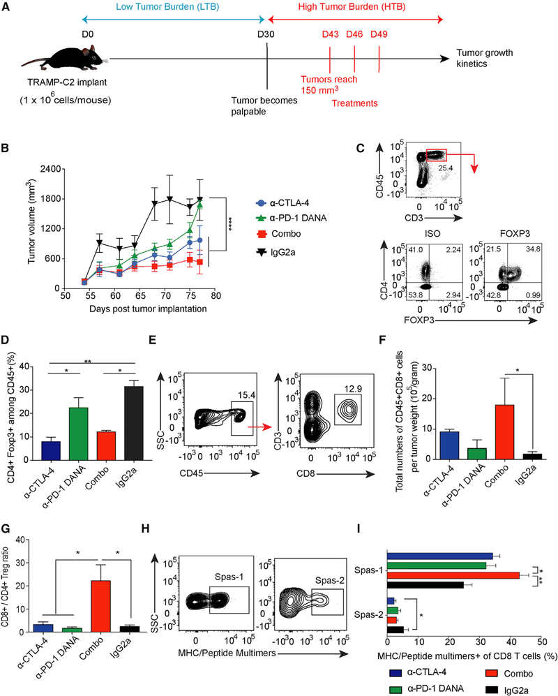
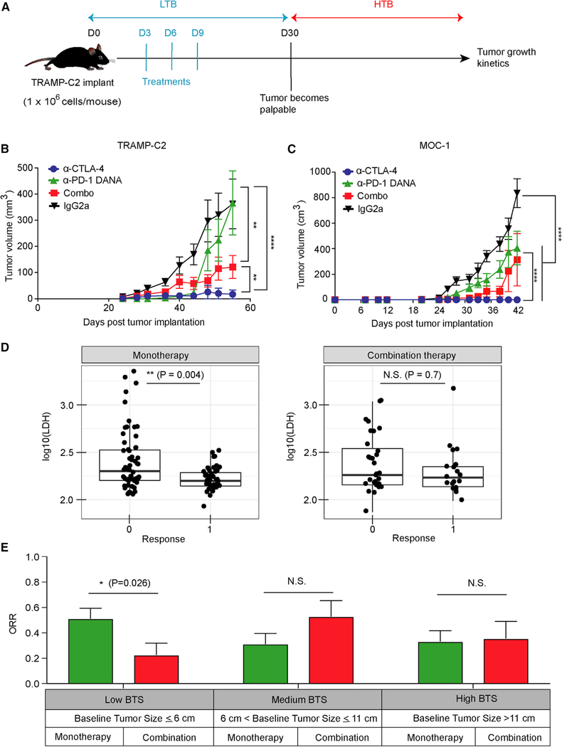
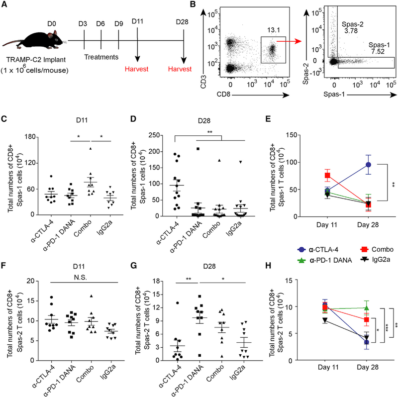
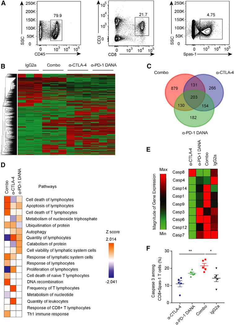
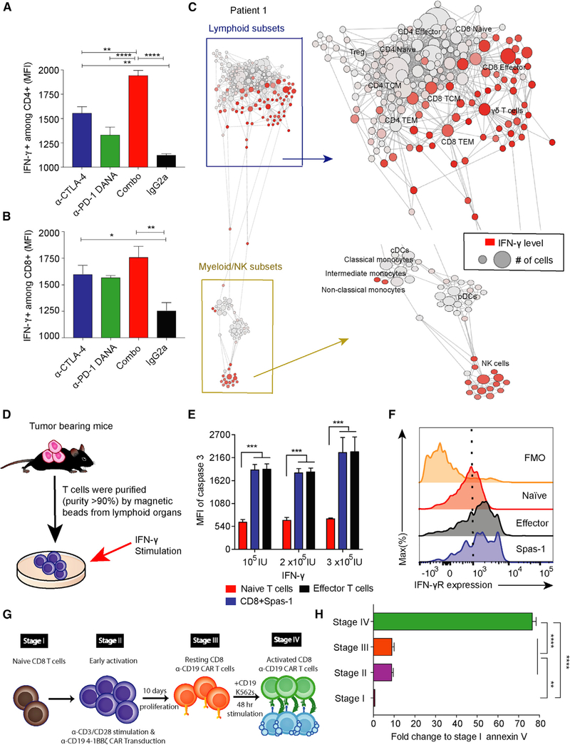
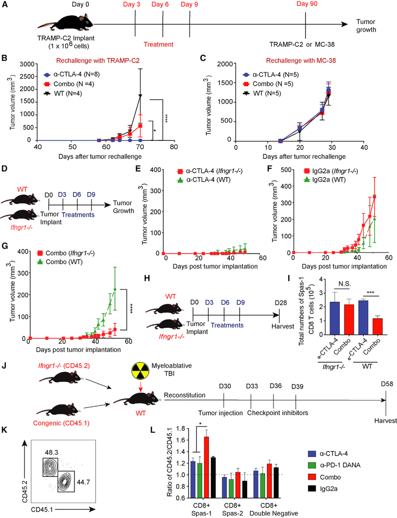
Resistance to checkpoint-blockade treatments is a challenge in the clinic. We found that although treatment with combined anti-CTLA-4 and anti-PD-1 improved control of established tumors, this combination compromised anti-tumor immunity in the low tumor burden (LTB) state in pre-clinical models as well as in melanoma patients. Activated tumor-specific T cells expressed higher amounts of interferon-γ (IFN-γ) receptor and were more susceptible to apoptosis than naive T cells. Combination treatment induced deletion of tumor-specific T cells and altered the T cell repertoire landscape, skewing the distribution of T cells toward lower-frequency clonotypes. Additionally, combination therapy induced higher IFN-γ production in the LTB state than in the high tumor burden (HTB) state on a per-cell basis, reflecting a less exhausted immune status in the LTB state. Thus, elevated IFN-γ secretion in the LTB state contributes to the development of an immune-intrinsic mechanism of resistance to combination checkpoint blockade, highlighting the importance of achieving the optimal magnitude of immune stimulation for successful combination immunotherapy strategies.

Keywords: IFN-γ; activation-induced cell death; anti-CTLA-4; anti-PD-1; cancer; immunotherapy.


Conflict of interest statement

DECLARATION OF INTERESTS

The other authors declare no conflicts of interest.

Figures

Figure 1.
Figure 1.. Combination Checkpoint Blockade Enhances Anti-tumor Responses against Established Tumors
C57BL/6j mice were implanted with TRAMP-C2 tumors and treated with checkpoint inhibitors as indicated. (A) Schema of mice injected with checkpoint blockade in the HTB state. (B) Tumor growth curve of the TRAMP-C2 model. (C) Flow staining of CD45+CD3+CD4+Foxp3+ T cells. (D) Percentage of CD4+Foxp3+ cells among CD45+ cells. (E) Flow gating strategy of CD8+ T cell subsets in tumors. (F) Total numbers of CD8+T cells per tumor weight. (G) Ratio of CD8+ to CD4+ Treg cells. (H and I) Tetramer staining among tumor-infiltrating CD8+ T cell populations. Cells were pre-gated on CD45+CD3+CD8+. Data were collected from at least eight mice per group with two independent experiments. Statistical differences were calculated by one-way or two-way ANOVA with a post hoc Tukey test. *p < 0.05, **p < 0.01, ****p < 0.0001. Data are presented as mean ± SE. See also Figure S1.
Figure 2.
Figure 2.. The Effect of Combination Checkpoint Blockade in the LTB State
(A) Schema of early intervention with dual checkpoint blockade prior to the development of palpable tumors. (B) TRAMP-C2 tumor growth with early intervention in different treatment groups. The dose for each checkpoint inhibitor was 10 mg/kg. (C) Tumor growth in the MOC-1 tumor model. The dose for each checkpoint inhibitor was 10 mg/kg. Animal studies were from two independent experiments with eight mice per group. Statistical analyses were calculated by two-way ANOVA with a post hoc test. *p < 0.05, **p < 0.01, ****p < 0.0001. (D) Comparison of LDH levels between patients treated with anti-PD-1 (monotherapy) and patients treated with anti-PD-1+anti-CTLA-4 (combination therapy). (E) 153 metastatic melanoma patients treated with checkpoint inhibitors were stratified into three groups with different ranges of baseline tumor size (BTS) as measured by radiographic imaging. The best overall response rate (RECIST v.1.1) in patients treated with monotherapy or combination therapy is presented for each stratum. Error bars represent SEM. Significance was calculated by the two-sided Mann-Whitney test. *p < 0.05. See also Figure S2.
Figure 3.
Figure 3.. Dynamics of Tumor-Specific T Cells after Checkpoint Blockade
Mice implanted with TRAMP-C2 tumors were treated with checkpoint inhibitors on days 3, 6, and 9. Spleens were harvested on days 11 and 28. (A) Schema of animal studies. (B) Flow gating of antigen-specific CD45+CD3+CD8+ T cells against the immunodominant Spas-1 epitope and minor Spas-2 epitope. (C) Total CD8+ Spas-1 T cells isolated at day 11. (D) Total CD8+ Spas-1 T cells isolated at day 28. (E) Dynamic changes of CD8+Spas-1 T cells over time. (F) Total CD8+ Spas-2 T cells isolated at day 11. (G) Total CD8+ Spas-2 T cells isolated at day 28. (H) Dynamic changes of CD8+ Spas-2 T cells over time. Data were from two or three independent experiments with 9–12 mice per group. Statistical analyses were calculated by one-way ANOVA with a post hoc Tukey test. *p < 0.05, **p < 0.01. Data are presented as mean ± SE. See also Figure S3.
Figure 4.
Figure 4.. Tumor-Specific T Cell Loss after Combination Checkpoint Inhibition
Mice were implanted with TRAMP-C2 tumors and treated with different checkpoint inhibitors. (A) CD8+ Spas-1 T cells were sorted from draining lymph nodes on day 28 after checkpoint-inhibitor treatment. (B) RNA-seq was performed on sorted cells, and expression data are presented for each treatment group. (C) The overlap in overexpressed genes was assessed for different treated groups. (D) Ingenuity Pathway Analysis was performed from RNA-seq data. (E) Gene expression for caspase family members was assessed by RT-PCR. (F) Active caspase-3 expression among CD8+ Spas-1 T cells was determined by flow cytometry. Data represent two independent experiments with ten mice per group. Statistical analyses were calculated by one-way ANOVA with post hoc Tukey test. *p < 0.05, **p < 0.01. Data are presented as mean ± SE. See also Figure S4.
Figure 5.
Figure 5.. IFN-γ Induced Cell Death of T Cells at Effector Stage
(A and B) Mice implanted with TRAMP-C2 tumors were treated with checkpoint inhibitors on days 3, 6, and 9. Spleens were harvested on day 11, and IFN-γ expression was analyzed in CD4+ and CD8+ T cell subsets by flow cytometry. (C) PBMCs were isolated from a total of nine cancer patients treated with anti-CTLA-4 plus anti-PD-1 therapies. Data were collected 1 month after the fourth dose of dual-checkpoint-blockade treatments. The data presented here were collected from patient 1, and the other eight patients are shown in Figure S5. Patient 1 is the same patient as reported in Figure S4. The IFN-γ secretion among different immune subsets was analyzed by CyTOF and is presented with SCAFFOLD. The node size represents the abundance of the cell population, and the red color represents the intensity of IFN-γ expression (ASINH ratio of the raw value). (D) T cells were purified from TRAMP-C2-tumor-bearing mice and subsequently cultured in vitro with the indicated concentrations of IFN-γ. (E) Cells were harvested 72 h after IFN-γ stimulation and analyzed for active caspase-3 expression among different CD8+ subsets. (F) IFN-γ receptor expression in different CD8+ T cell subsets. (G) Four stages of human chimeric antigen receptor (CAR) CD8+ T cell differentiation in vitro are shown. (H) CD8+ T cells at these different stages of activation were cultured in vitro with human recombinant IFN-γ (3 × 10⁵ IU), and cells were harvested 48 h later. Annexin V expression was determined by flow cytometry. Statistical analyses were calculated by one-way ANOVA with a post hoc Tukey test. **p < 0.01, ****p < 0.0001. Data are presented as mean ± SE. See also Figure S5.
Figure 6.
Figure 6.. Effective Anti-tumor Response with Dual Checkpoint Blockade Is Restored when T Cells Are Made Unresponsive to IFN-γ
(A) Eight-week-old C57BL/6j mice were challenged with TRAMP-C2 tumor on day 0 and treated with checkpoint inhibitors on days 3, 6, and 9. Ninety days after tumor implantation, tumor-free mice from CTLA-4 blockade or combination-treatment groups were subsequently re-challenged with either the TRAMP-C2 or MC-38 tumor model. Aged sibling mice without prior tumor challenge were used as control mice. (B) Mice were re-challenged with TRAMP-C2 tumors. (C) Mice were re-challenged with MC-38 tumors. To allow a sufficient number of tumor-free mice, each treatment group consisted of 30–45 mice. The numbers of tumor-free mice for re-challenge are labeled correspondingly. (D) Age-and gender-matched WT or Ifngr1−/− C57BL/6j mice were implanted with tumors at day 0 and treated with checkpoint inhibitors on days 3, 6, and 9. (E) Comparison of tumor growth curves in the CTLA-4-blockade treatment. (F) Comparison of tumor growth curves in the isotype-control treatment. (G) Comparison of tumor growth curves with the combination-blockade treatment. (H) WT or Ifngr1−/− mice were treated with checkpoint blockade and harvested at day 28. (I) Total numbers of CD8+ Spas-1 T cells. (J) WT mice were myeloablated (10.5 Gy) and given an adoptive transfer of bone marrow cells from CD45.2 Ifngr1−/− and CD45.1 Pepc congenic WT mice at a 1:1 ratio. Chimera mice were subsequently implanted with TRAMP-C2 tumors and treated with checkpoint inhibitors on days 33, 36, and 39 after bone marrow transplant. (K) Mice underwent tail bleeding and were checked for chimerism 30 days after bone marrow transplantation. (L) Tumor-draining lymph nodes were harvested on day 58. Different CD8+ subsets were pre-gated on flow cytometry and investigated for chimerism. Experiments included eight (D–G) or five (H and I) mice per group. For (J) and (L), each treatment consisted of five chimera mice per group for a total of 20 chimera mice in these experiments. Data were analyzed by two-way ANOVA with a post hoc test (B–G) or by one-way ANOVA with a post hoc Tukey test (I and L). *p < 0.05, ***p < 0.001, ****p < 0.0001. Data are presented as mean ± SE. See also Figure S6.

Comment in

References

    1. Badovinac VP, Tvinnereim AR, and Harty JT (2000). Regulation of antigen-specific CD8+ T cell homeostasis by perforin and interferon-gamma. Science 290, 1354–1358. - PubMed
    1. Bellmunt J, de Wit R, Vaughn DJ, Fradet Y, Lee JL, Fong L, Vogelzang NJ, Climent MA, Petrylak DP, Choueiri TK, et al. ; KEYNOTE-045 Investigators (2017). Pembrolizumab as second-line therapy for advanced urothelial carcinoma. N. Engl. J. Med. 376, 1015–1026. - PMC - PubMed
    1. Benci JL, Xu B, Qiu Y, Wu TJ, Dada H, Twyman-Saint Victor C, Cucolo L, Lee DSM, Pauken KE, Huang AC, et al. (2016). Tumor interferon signaling regulates a multigenic resistance program to immune checkpoint blockade. Cell 167, 1540–1554.e12. - PMC - PubMed
    1. Berner V, Liu H, Zhou Q, Alderson KL, Sun K, Weiss JM, Back TC, Longo DL, Blazar BR, Wiltrout RH, et al. (2007). IFN-gamma mediates CD4+ T-cell loss and impairs secondary antitumor responses after successful initial immunotherapy. Nat. Med. 13, 354–360. - PubMed
    1. Borghaei H, Paz-Ares L, Horn L, Spigel DR, Steins M, Ready NE, Chow LQ, Vokes EE, Felip E, Holgado E, et al. (2015). Nivolumab versus docetaxel in advanced nonsquamous non-small-cell lung cancer. N. Engl. J. Med. 373, 1627–1639. - PMC - PubMed

Publication types

MeSH terms