. 2019 Apr 1;202(7):2035-2043.
doi: 10.4049/jimmunol.1801161. Epub 2019 Feb 8.

Elevated Expression of Macrophage Migration Inhibitory Factor Promotes Inflammatory Bone Resorption Induced in a Mouse Model of Periradicular Periodontitis

Affiliations

Elevated Expression of Macrophage Migration Inhibitory Factor Promotes Inflammatory Bone Resorption Induced in a Mouse Model of Periradicular Periodontitis

Mohammed Howait et al. J Immunol. .

Abstract

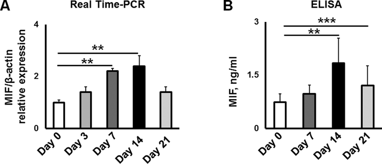
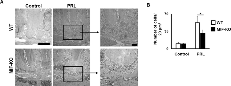
Locally produced osteoclastogenic factor RANKL plays a critical role in the development of bone resorption in periradicular periodontitis. However, because RANKL is also required for healthy bone remodeling, it is plausible that a costimulatory molecule that upregulates RANKL production in inflammatory periradicular periodontitis may be involved in the pathogenic bone loss processes. We hypothesized that macrophage migration inhibitory factor (MIF) would play a role in upregulating the RANKL-mediated osteoclastogenesis in the periradicular lesion. In response to pulp exposure, the bone loss and level of MIF mRNA increased in the periradicular periodontitis, which peaked at 14 d, in conjunction with the upregulated expressions of mRNAs for RANKL, proinflammatory cytokines (TNF-α, IL-6, and IL-1β), chemokines (MCP-1 and SDF-1), and MIF's cognate receptors CXCR4 and CD74. Furthermore, expressions of those mRNAs were found significantly higher in wild-type mice compared with that of MIF-/- mice. In contrast, bacterial LPS elicited the production of MIF from ligament fibroblasts in vitro, which, in turn, enhanced their productions of RANKL and TNF-α. rMIF significantly upregulated the number of TRAP+ osteoclasts in vitro. Finally, periapical bone loss induced in wild-type mice were significantly diminished in MIF-/- mice. Altogether, the current study demonstrated that MIF appeared to function as a key costimulatory molecule to upregulate RANKL-mediated osteoclastogenesis, leading to the pathogenically augmented bone resorption in periradicular lesions. These data also suggest that the approach to neutralize MIF activity may lead to the development of a therapeutic regimen for the prevention of pathogenic bone loss in periradicular periodontitis.


Figures

Figure 1.
Figure 1.
Time course analysis of MIF mRNA expressions (A) and protein concentrations (B) in mouse periradicular tissues. MIF gene expression was evaluated by Real-Time PCR at Day 3, 7, 14 and 21 after pulp exposure. Protein levels were checked by ELISA at Days 7, 14 and 21 after pulp exposure. **p<0.01, ***p<0.001
Figure 2.
Figure 2.
Fibroblasts are one of the major source of MIF in a mouse model of periradicular lesion. A: Expression of MIF mRNA by primary human periodontal ligament fibroblasts. Periodontal ligament fibroblasts were stimulated with various concentrations of E. coli-LPS, S. aureus-LTA, and pro-inflammatory cytokines (recombinant TNF-α and IL-1β proteins). B: MIF production by primary human periodontal ligament fibroblasts in the presence of LPS. Periodontal ligament fibroblasts were stimulated with various concentrations of E. coli-LPS, S. aureus-LTA, and pro-inflammatory cytokines (recombinant TNF-α and IL-1β proteins). The collected mRNA and supernatants were analyzed by Real Time-PCR and ELISA after 12 h and 24h of stimulation, respectively. C: MIF co-localization with ligament fibroblasts in healthy control and diseased periradicular tissue at Day 14 after pulp exposure. White dash line represents transition in MIF (red) expression between non-inflamed periodontal ligament (PDL) and periradicular lesion (PRL). Fibroblasts and nuclei are stained with anti-PDL-1 Ab (green) and DAPI (blue), respectively. Scale bar = 50 μm. *p<0.05, ** p<0.01, *** p<0.001.
Figure 3.
Figure 3.
Effect of anti-MIF neutralizing mAb on production of osteoclastogenic RANKL (A) and pro-inflammatory cytokine TNF-α (B) from mouse gingival fibroblasts in response to LPS stimulation. Cell were stimulated with LPS isolated from E. coli (10 ng/ml) in the presence of either anti-MIF mAb or control-mAb (1 μg/ml of each). After 24 h of stimulation, the amounts of RANKL and TNF-α in collected supernatant were evaluated by ELISA. * p<0.05, ** p<0.01, *** p<0.001.
Figure 4.
Figure 4.. Expressions of MIF’s cognate receptors, CD74 and CXCR4, on LPS-stimulated fibroblasts.
Mouse gingival fibroblasts were stimulated with E. coli-LPS (10 ng/ml) in the presence of either anti-MIF mAb or control-mAb (1 μg/ml of each). After 24 h of stimulation, the expression of CD74 and CXCR4 were evaluated by multicolor flow cytometry. * p<0.05, *** p<0.001.
Figure 5.
Figure 5.
Expression of the mRNAs for CD74 (A) and CXCR4 (B) in periradicular lesions (PRL) induced in WT and MIF−/− mice. The periradicular tissue was collected at Day 14 after the pulp exposure. N= 5 mice/condition. The Student’s t-test was used to evaluate statistically significant differences. A.U – arbitrary units. *** p<0.001.
Figure 6.
Figure 6.. Expression patterns of mRNA for pro-inflammatory cytokines and chemokines, and RANKL in the periradicular lesions induced in WT mice vs MIF−/− mice.
The expressions of mRNA for pro-inflammatory cytokines IL-1b, IL-6, and TNF-a (A) and chemokines MCP-1 and SDF-1 (B), and RANKL (C) in WT and MIF−/− mice in the periradicular lesions (PRL). ** p<0.01, *** p<0.001.
Figure 7.
Figure 7.
Histological evaluation of mononuclear cell infiltrations to periradicular lesions (PRL) induced in MIF−/− and WT mice. A: Evaluation of histo-morphological changes in periradicular tissue induced by pulp exposure of third molar at Day 14 in WT and MIF−/− mice. B: The number of recruited cells quantified from the histological section (A). Scale bar = 20 μm. * p<0.05
Figure 8.
Figure 8.
Effects of recombinant MIF on RANKL-induced osteoclastogenesis in vitro. Microscopic evaluation of the TRAP+ and quantification of the number of TRAP+ cells in Raw 264.7 cells (A & B) and bone marrow derived macrophages (C & D). E & F: Microscopic evaluation and quantification of the pit formation area. RANKL-stimulated RAW264.7 cells and bone marrow derived macrophages (in the presence of M-CSF) were incubated with various concentrations of mouse recombinant MIF protein. The cells are evaluated at day 5 after stimulation. * p<0.05, ** p<0.01, *** p<0.001.
Figure 9.
Figure 9.
Manifestations of pathologic bone loss phenotypes found in periradicular lesion induced in MIF−/− compared to WT mice. μCT images of murine periradicular lesions (A) and quantitative measurement of bone resorption area (B) in WT and MIF−/− mice evaluated 14 days after third molar exposure. Histological evaluation of TRAP+ osteoclasts (C) measured in a microscopic field of TRAP-stained sections (D) in WT and MIF−/− mice. A single section through each mandible is shown and arrows indicate the areas of bone loss. *p<0.05, ** p<0.01.

References

    1. Marmary Y, and Kutiner G (1986) A radiographic survey of periapical jawbone lesions. Oral surgery, oral medicine, and oral pathology 61, 405–408 - PubMed
    1. Lin LM, Ricucci D, Lin J, and Rosenberg PA (2009) Nonsurgical root canal therapy of large cyst-like inflammatory periapical lesions and inflammatory apical cysts. Journal of endodontics 35, 607–615 - PubMed
    1. Nair PN (1997) Apical periodontitis: a dynamic encounter between root canal infection and host response. Periodontology 2000 13, 121–148 - PubMed
    1. Kawashima N, Okiji T, Kosaka T, and Suda H (1996) Kinetics of macrophages and lymphoid cells during the development of experimentally induced periapical lesions in rat molars: a quantitative immunohistochemical study. Journal of endodontics 22, 311–316 - PubMed
    1. Liapatas S, Nakou M, and Rontogianni D (2003) Inflammatory infiltrate of chronic periradicular lesions: an immunohistochemical study. International endodontic journal 36, 464–471 - PubMed

Publication types

MeSH terms

Substances