. 2019 Feb 22;39(2):BSR20181833.
doi: 10.1042/BSR20181833. Print 2019 Feb 28.

Metabolomic analysis reveals a protective effect of Fu-Fang-Jin-Qian-Chao herbal granules on oxalate-induced kidney injury

Affiliations

Metabolomic analysis reveals a protective effect of Fu-Fang-Jin-Qian-Chao herbal granules on oxalate-induced kidney injury

Wei Chen et al. Biosci Rep. .

Abstract

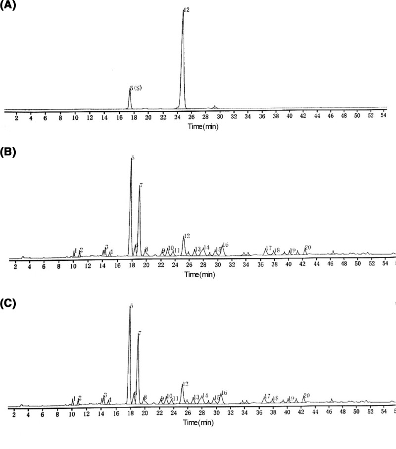
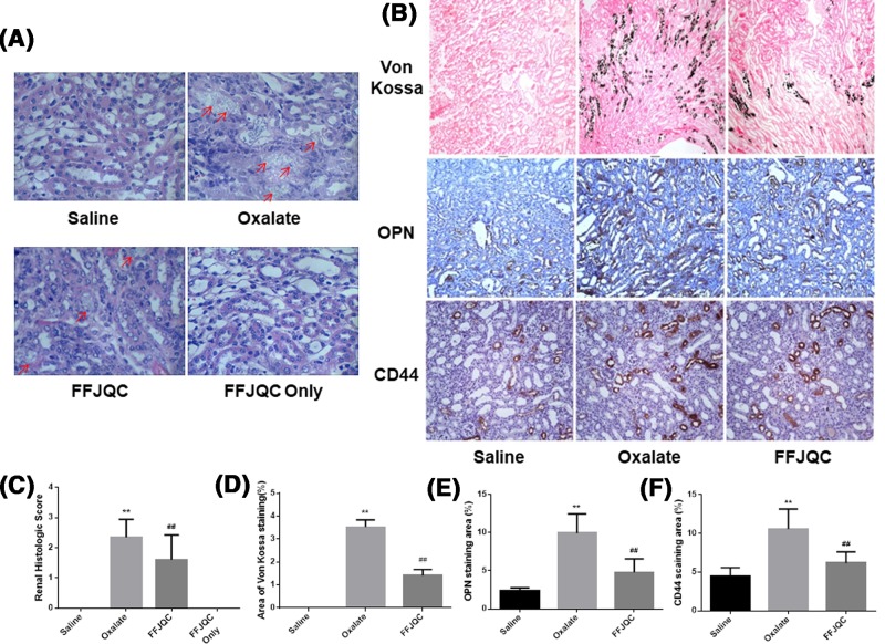
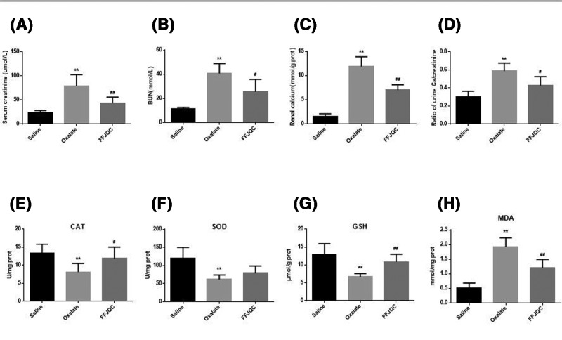
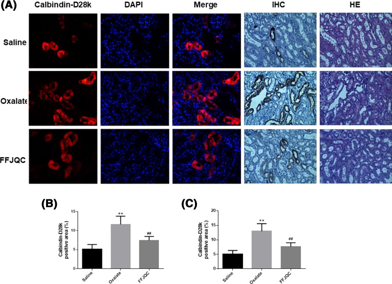
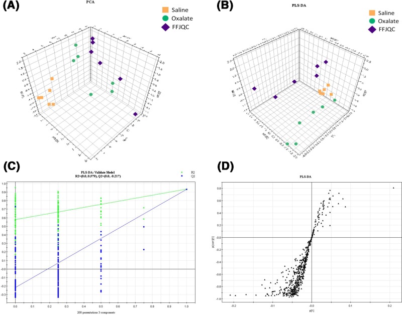
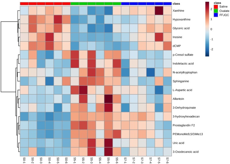
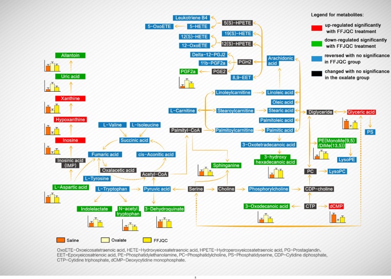
Nephrolithiasis is one of the world's major public health burdens with a high incidence and a risk of persistent renal dysfunction. Fu-Fang-Jin-Qian-Chao granules (FFJQC), a traditional Chinese herb formula, is commonly used in treatment of nephrolithiasis. However, the therapeutic mechanism of FFJQC on kidney stone has still been a mystery. The objective of the present study is to explore the therapeutic mechanism of FFJQC on kidney injury and identify unique metabolomics patterns using a mouse model of kidney stone induced by a calcium oxalate (CaOx) deposition. Von Kossa staining and immuno-histopathological staining of osteopontin (OPN), cluster of differentiation 44 (CD44) and calbindin-D28k were conducted on renal sections. Biochemical analysis was performed on serum, urine, and kidney tissues. A metabolomics approach based on ultra-HPLC coupled with quadrupole-TOF-MS (UHPLC-Q-TOF/MS) was used for serum metabolic profiling. The immunohistopathological and biochemical analysis showed the therapeutic benefits of FFJQC. The expression levels of OPN and CD44 were decreased while calbindin-D28k increased after the CaOx injured mice were treated with FFJQC. In addition, total of 81 serum metabolites were identified to be associated with protective effects of FFJQC on CaOx crystal injured mice. Most of these metabolites were involved in purine, amino acid, membrane lipid and energy metabolism. Potential metabolite biomarkers were found for CaOx crystal-induced renal damage. Potential metabolite biomarkers of CaOx crystal-induced renal damage were found. FFJQC shows therapeutic benefits on CaOx crystal injured mice via regulation of multiple metabolic pathways including amino acids, purine, pyrimidine, glycerolipid, arachidonic acid (AA), sphingolipid, glycerophospholipid, and fatty acid.

Keywords: Fu-Fang-Jin-Qian-Chao; Kidney stone; Oxalate crystal; mass spectrometry; metabolomics.


Conflict of interest statement

The authors declare that there are no competing interests associated with the manuscript.

Figures

Figure 1
Figure 1. High performance liquid chromatograms of FFJQC and the reference standards
(A) Chromatogram of standards with mangiferin(5) and schaftoside(12). (B,C) Chromatograms of two batches of FFJQC provided by Guangxi Wantong Pharmaceutical Co., Ltd. (180627/1 and 180705/1).
Figure 2
Figure 2. Histological analysis of mouse model induced by oxalate
(A) Representative images of HE staining of kidney cortex and medulla junction. (B) Representative photomicrographs of Von Kossa staining of calcium deposition and immunostaining of OPN and CD44 expression. The crystal deposition was clearly increased in the oxalate mouse group. After the FFJQC treatment, the crystal deposition was decreased significantly in this group. The protein expression levels of OPN and CD44 were increased in the oxalate group, then their levels were decreased significantly after the FFJQC treatment. (C) Tubulointerstitial damage were assessed semiquantitatively according to Carraway’s scoring from every 20 random views. (D) Semi-quantitative analysis of calcium deposition in the area of positive staining from 20 random views. (E,F) Semi-quantitative analysis of OPN and CD44 expression in each group from 20 random immunohistochemical views. (C) **P<0.0001 Oxalate group compared with Saline group, ##P=0.002 FFJQC group compared with Oxalate group. (D) **P<0.0001 Oxalate group compared with Saline group, ##P<0.0001 FFJQC group compared with Oxalate group. (E) **P<0.0001 Oxalate group compared with Saline group, ##P<0.0001 FFJQC group compared with Oxalate group. (F) **P<0.0001 Oxalate group compared with Saline group, ##P<0.0001 FFJQC group compared with Oxalate group.
Figure 3
Figure 3. Biochemical analysis of mouse model induced by oxalate
Levels of Scr (A), BUN (B), renal calcium (C), and urine Ca/creatinine (D) were determined (**P<0.01 oxalate group compared with saline group, #P<0.05, ##P<0.01 FFJQC group compared with oxalate group). All data are expressed as the mean ± S.D. (E) Concentration of CAT in the kidney, (F) concentration of SOD in the kidney, (G) concentration of GSH in the kidney, (H) concentration of MDA in the kidney. (A) **P=0.0002 Oxalate group compared with Saline group, ##P=0.0081 FFJQC group compared with Oxalate group; (B) **P<0.0001 Oxalate group compared with Saline group, #P=0.0177 FFJQC group compared with Oxalate group; (C) **P<0.0001 Oxalate group compared with Saline group, ##P=0.0004 FFJQC group compared with Oxalate group. (D) **P<0.0001 Oxalate group compared with Saline group, ##P=0.0139 FFJQC group compared with Oxalate group; (E) **P=0.0005 Oxalate group compared with Saline group, ##P=0.0447 FFJQC group compared with Oxalate group; (F) **P=0.0014 Oxalate group compared with Saline group, ##P=0.0947 FFJQC group compared with Oxalate group; (G) **P=0.0007 Oxalate group compared with Saline group, ##P=0.0014 FFJQC group compared with Oxalate group; (H) **P<0.0001 Oxalate group compared with Saline group, ##P=0.0019 FFJQC group compared with Oxalate group.
Figure 4
Figure 4. Representative immunofluorescent(IF) and immunohistochemical (IHC) staining of calbindin-D28k at the corticomedullary junction in kidneys
The calbindin-D28k protein were labeled with red fluorescence and the nuclei were labeled with blue fluorescence. The images were taken under fluorescence microscope-Magnification: 400×, optical microscope-Magnification: 200×. (A) The calbindin-D28k expression level decreased in the oxalate mouse group. After the FFJQC treatment, the calbindin-D28k expression level increased significantly. (B) Quantitation of calbindin-D28k+ area in IF, **P<0.001 Oxalate group compared with Saline group, ##P=0.0015 FFJQC group compared with Oxalate group. (C) Quantitation of calbindin-D28k+ area in IHC, **P<0.001 Oxalate group compared with Saline group, ##P=0.0021 FFJQC group compared with Oxalate group. Twenty specimens were used while calculating the calbindin-positive area.
Figure 5
Figure 5. Plots of multivariate statistical analysis based on the serum metabolites in ESI positive ion mode
(A) PCA scores plot of saline, oxalate, and FFJQC groups. (B) PLS-DA scores plot of saline, oxalate, and FFJQC groups. (C) Validation plot obtained from permutation tests. (D) S-plot of the PLS-DA model.
Figure 6
Figure 6. Heatmap generated from the relative levels of key metabolite biomarkers in mouse serum from groups of saline, oxalate, and FFJQC treated
Figure 7
Figure 7. Proposed model of metabolic pathway networks resulting from the glyoxylate-induced crystal kidney injury and FFJQC treatment

References

    1. Sorokin I., Mamoulakis C., Miyazawa K., Rodgers A., Talati J. and Lotan Y. (2017) Epidemiology of stone disease across the world. World J. Urol. 35, 1–20 10.1007/s00345-017-2008-6 - DOI - PubMed
    1. Lee Y.C., Huang S.P., Juan Y.S., Huang T.Y. and Liu C.C. (2016) Impact of metabolic syndrome and its components on kidney stone in aging Taiwanese males. Aging Male 19, 197–201 10.1080/13685538.2016.1174987 - DOI - PubMed
    1. Sáenzmedina J., Jorge E., Corbacho C., Santos M., Sánchez A., Soblechero P.. et al. (2017) Metabolic syndrome contributes to renal injury mediated by hyperoxaluria in a murine model of nephrolithiasis. Urolithiasis 46, 1–8 - PubMed
    1. Favus M.J. (2011) The risk of kidney stone formation: the form of calcium matters. Am. J. Clin. Nutr. 94, 270–277 10.3945/ajcn.111.018481 - DOI - PubMed
    1. Alexander R.T., Hemmelgarn B.R., Wiebe N., Bello A., Morgan C., Samuel S.. et al. (2012) Kidney stones and kidney function loss: a cohort study. BMJ 345, e5287 10.1136/bmj.e5287 - DOI - PMC - PubMed

Publication types