Review
. 2019 Mar;20(3):148-160.
doi: 10.1038/s41583-019-0132-6.

Oxidative stress, dysfunctional glucose metabolism and Alzheimer disease

Affiliations
Review

Oxidative stress, dysfunctional glucose metabolism and Alzheimer disease

D Allan Butterfield et al. Nat Rev Neurosci. 2019 Mar.

Abstract

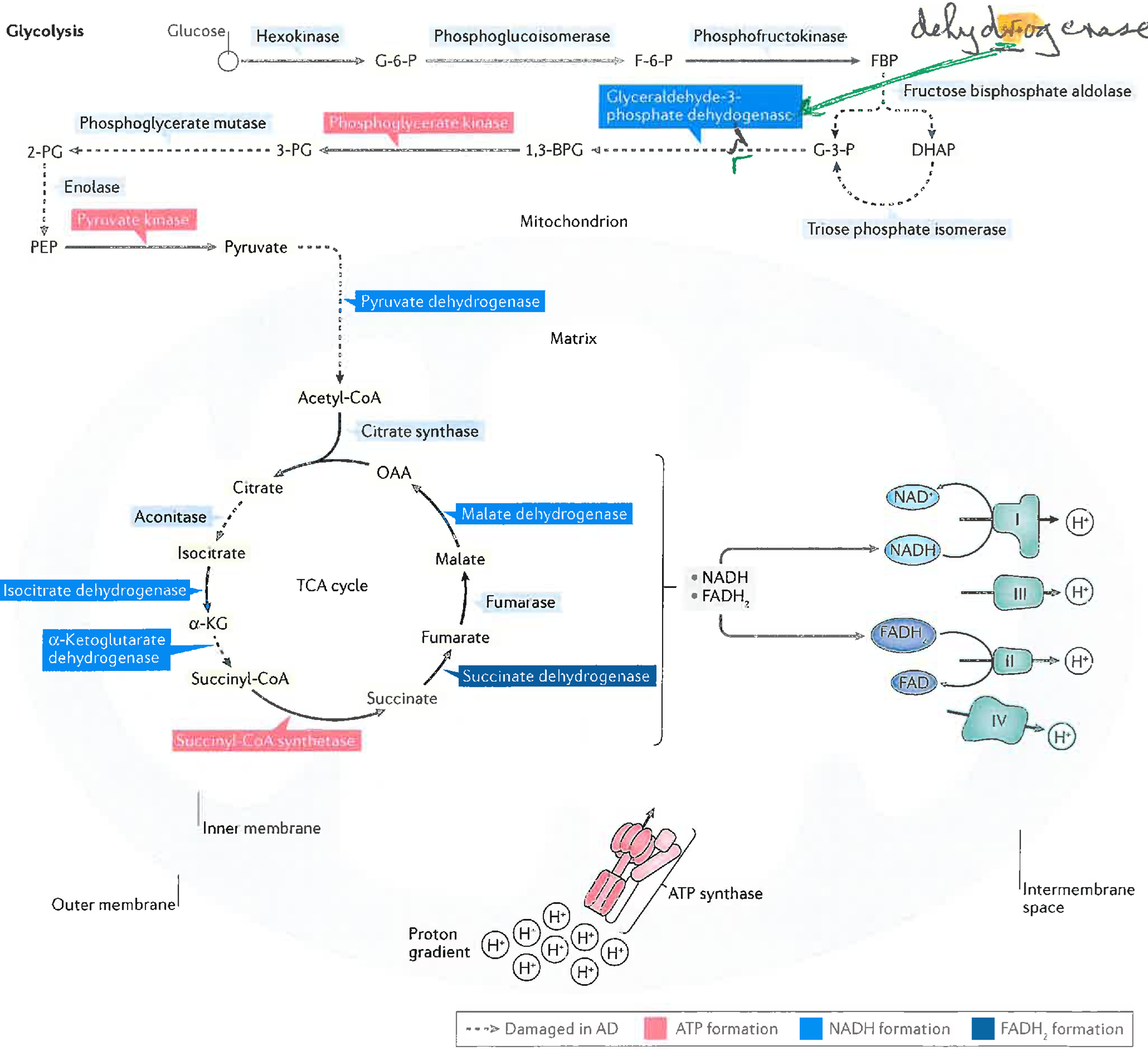
Alzheimer disease (AD) is a major cause of age-related dementia. We do not fully understand AD aetiology and pathogenesis, but oxidative damage is a key component. The brain mostly uses glucose for energy, but in AD and amnestic mild cognitive impairment glucose metabolism is dramatically decreased, probably owing, at least in part, to oxidative damage to enzymes involved in glycolysis, the tricarboxylic acid cycle and ATP biosynthesis. Consequently, ATP-requiring processes for cognitive function are impaired, and synaptic dysfunction and neuronal death result, with ensuing thinning of key brain areas. We summarize current research on the interplay and sequence of these processes and suggest potential pharmacological interventions to retard AD progression.


Conflict of interest statement

Competing interests

Competing interests policy There is NO Competing Interest.

Figures

Figure 1.
Figure 1.. Schematic diagrams of the biochemistry of glucose catabolism and ATP synthesis and their oxidative dysfunction in AD and aMCI brains
Glycolysis, the tricarboxylic acid (TCA) cycle, and the electron transport chain (ETC), the latter localized on the inner mitochondrial membrane, work together to catabolize glucose and drive ATP synthesis via the ATP synthase complex. Complexes I to IV of the ETC are shown. Also shown is ATP Synthase, whose α-chain is oxidatively modified in brains of subjects with AD. Briefly, this figure shows that glucose is converted to pyruvate in glycolysis. Pyruvate is converted to acetyl coenzyme A, which enters the TCA cycle, and resulting reducing equivalents (NADH and FADH2) from glycolysis and the TCA cycle enter the mitochondrial ETC. (The inner mitochondrial membrane is impermeable to NADH, so the Malate-Aspartate shuttle leads to NADH synthesis in the matrix via NADH in the cytosol) to reduce oxygen to water, leading to production of a mitochondrial proton gradient in the Intermembrane Space that drives ATP synthesis. Reactions catalyzed by specific enzymes or enzyme complexes identified by redox proteomics or other techniques to be oxidatively damaged (and likely thereby dysfunctional) in AD brain (and most also in aMCI brains),,–,,, are indicated as dashed lines in the Figure. Abbreviations: G-6-P, glucose-6-phosphate; F-6-P, fructose-6-phosphate; FBP, fructose-1,6-bisphosphate; DHAP, dihydroxyacetone phosphate; G-3-P, glyceraldehyde-3-phosphate; 1,3-BPG, 1,3-bisphosphoglycerate; 3-PG, 3-phosphoglycerate; 2-PG, 2-phosphoglycerate; PEP, phosphoenolpyruvate; acetyl-CoA, acetyl coenzyme A; α-KG, α-ketoglutarate; succinyl-CoA, succinyl-coenzyme A; OAA, oxaloacetate.
Figure 2.
Figure 2.. Schematic representation of biochemical events associated with insulin binding to its receptor, leading to activation of mTORC1 with subsequent inhibition of autophagy and development of insulin resistance
After insulin (INS) binds to the insulin receptor (IR) on neuronal membranes, the IR dimerizes and auto-phosphorylation on tyrosine residues occurs. The insulin receptor substrate-1 (IRS-1) recognizes IR phosphotyrosine residues and binds to the IR, which in turn leads to phosphorylation of IRS-1 on tyrosine residues 612 and 632 with resultant activation of IRS-1. Activated IRS-1 leads to phosphorylation and activation of two pathways for the insulin signaling cascade, one of which is the PI3/Akt pathway. Phosphorylated PI3K leads to phosphorylation and activation of Akt, which leads to phosphorylation of Ser-2448 of the mechanistic target of rapamycin complex 1 (mTORC1), the latter becoming activated as a kinase,,,. Activated mTORC1 kinase has several key downstream effects (two of which are shown in the figure) that impair neuronal survival (and are thus relevant to AD). There are (a) inhibition of autophagy; and (b) phosphorylation of the protein, p70S6K, which then becomes a kinase, one of whose substrates is Ser-307 of IRS-1. Once phosphorylated on Ser-307, IRS-1 function ceases, leading to and becoming a marker for insulin resistance,,. Modified with permission from Ref. .
Figure 3.
Figure 3.. Schematic drawings of the three components of the proteostasis network in brain cells.
A: The Ubiquitin Proteasomal System (UPS). Damaged proteins are polyubiquitinylated by the Ubiquitin Ligase Enzymes, E1, E2, E3. E1 requires ATP for its function. An initial ubiquitin (Ub) molecule is bound to the damaged protein or organelle by this process and is repeated to form a polyubiquitinylated chain. Polyubiquitinylated damaged proteins are destined for degradation by the 26S proteasome, but prior to entering the 19S cap, these proteins must be de-ubiquitinylated, one ubiquitin residue at a time by the enzyme, ubiquitin carboxyl-terminal hydrolase L-1 (UCH L1). The de-ubiquitinylated, damaged protein is degraded by the proteinases in the 20S portion of the 26S proteasome, and small peptides are ejected by the bottom 19S portion of the 26S proteasome to become degraded by soluble peptidases to individual amino acids for reuse. B: Autophagic Degradation of Aggregated Proteins or Organelles. The process starts with formation of a double membrane enveloping the aggregated, damaged protein or organelle, to form an autophagosome. This is transported to the lysosome, where membrane fusion leads to formation of the autophagolysosome. Endocytosis of the contents of the autophagosome into the acidic interior of the lysosome leads to their proteolytic degradation, with peptides, amino acids, and other biomolecules being ejected from the autophagolysosome for reuse. C: The Unfolded Protein Response (UPR) Associated with ER Stress. Following an elevation in the levels of misfolded proteins and/or Ca2+ in the ER lumen, the UPR is usually engaged. This consists of activation of one or more of three stress transducers: protein kinase R-like ER kinase (PERK), inositol-requiring enzyme 1alpha (IRE1), and activating transcription factor-6 alpha and beta (ATF6). Activation of each stress sensor is accompanied by removal from the stress sensor of binding immunoglobulin protein (BiP, also known as glucose regulated protein 78, Grp78), which when bound inactivates each of the three stress transducers. Each activated stress sensor induces one or more down-stream response mechanisms. In the figure, up arrows denote increased process or level, whereas down arrows denote decreased process or level. For PERK, receptor Tyr-kinase activity phosphorylates eIF2α that leads to decreased protein translation, which causes elevated rates of translation of normally poorly translated mRNAs, among which is ATF4. This, in turn, leads to decreased redox homeostasis and elevated apoptosis. ATF6 transduces ER stress by inducing transport of the ATF6 precursor protein to the Golgi apparatus, where the shorter ATF6 is produced. The latter translocates to the nucleus leading to expression of X-box binding protein (XBP1). In the case of IRE1, transduced ER stress results in dimerization and formation of a transmembrane kinase – Receptor Tyr Kinase (RTK), which in turn results in: a) RNA degradation by regulated IRE1-dependent decay (RIDD); b) XBP1-mediated alternative mRNA splicing that leads to ER-associated degradation (ERAD) and increased lipid metabolism (from which elevated levels of the lipid peroxidation product, 4-hydroxynonenal (HNE) can arise); and c) tumor necrosis factor receptor-associated factor 2 (TRAF2)-mediated inflammatory or pro-apoptotic gene induction, particularly those of nuclear factor kappa-light chain enhancer of activated B cells (NF-κB) and c-Jun N-terminal kinase (JNK). The endoplasmic reticulum (ER)-resident component of the proteostasis network, particularly the UPR, is impaired in aMCI and AD brain, due to accumulation of abnormal proteins and alterations in Ca2+ homeostasis–,. In AD brain, markers of ER stress are elevated and correlate with progression of the disease–,. Modified with permission from Ref. .

References

    1. Nelson PT, Braak H & Markesbery WR Neuropathology and cognitive impairment in Alzheimer disease: a complex but coherent relationship. J. Neuropathol. Exp. Neurol 68,1–14 (2009). - PMC - PubMed
    1. Martins RN et al. Alzheimer’s disease: a journey from amyloid peptides and oxidative stress, to biomarker technologies and disease prevention strategies—gains from AIBL and DIAN cohort studies. J. Alzheimers Dis 62, 965–992 (2018). - PMC - PubMed
    2. A detailed review of how cohort studies have contributed to our understanding of AD

    1. Markesbery WR Neuropathologic alterations in mild cognitive impairment: a review. J. Alzheimers Dis 19, 221–228 (2010). - PMC - PubMed
    1. Landau SM & Frosch MP Tracking the earliest pathological changes in Alzheimer disease. Neurology 82, 878–883 (2014). - PubMed
    1. Weise CM et al. Left lateralized cerebral glucose metabolism declines in amyloid-β positive persons with mild cognitive impairment. Neuroimage. Clin 20, 286–296 (2018). - PMC - PubMed

Publication types