Review
. 2019 Mar;26(4):715-727.
doi: 10.1038/s41418-019-0297-6. Epub 2019 Feb 8.

Regulation of the innate immune system by autophagy: monocytes, macrophages, dendritic cells and antigen presentation

Affiliations
Review

Regulation of the innate immune system by autophagy: monocytes, macrophages, dendritic cells and antigen presentation

Nina Germic et al. Cell Death Differ. 2019 Mar.

Abstract

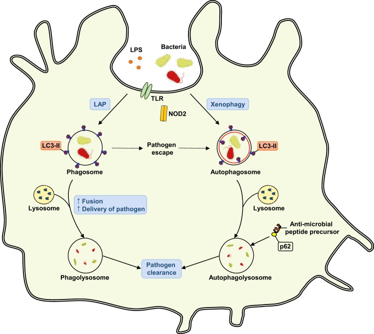
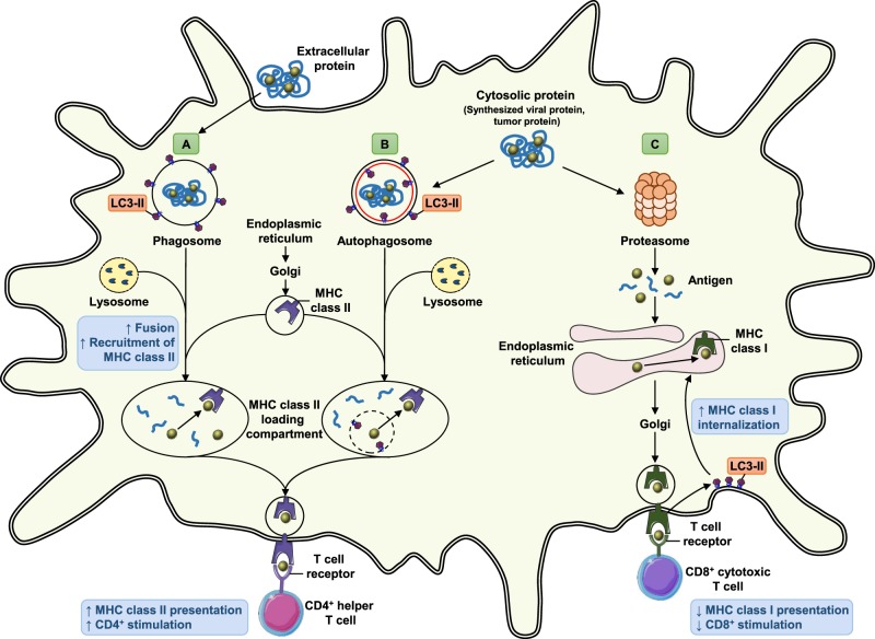
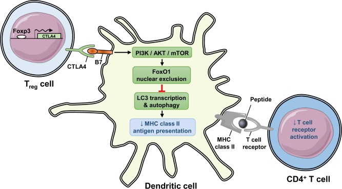
Autophagy is well equipped functionally to isolate microbial pathogens in autophagosomes and to carry out their clearance by dismemberment in the course of catabolic processes in the lysosome. Clearly, this is a non-metabolic function of autophagy that impacts strongly on the immune system. While in a preceding article on neutrophils, eosinophils, mast cells, and natural killer cells our focus was on the role of autophagy in regulating innate immune cell differentiation, degranulation, phagocytosis and extracellular trap formation, here we discuss monocytes/macrophages and dendritic cells, specifically, the influence of autophagy on functional cellular responses, such as phagocytosis, antigen presentation, cytokine production, control of inflammasome activation, tolerance and the consequences for overall host defense.


Conflict of interest statement

The authors declare that they have no conflict of interest.

Figures

Fig. 1
Fig. 1
Autophagic activity in the differentiation from monocytes to macrophages. Innate immune cells such as macrophages and DCs arise from a hematopoietic stem cell (HSC) that has committed to a common myeloid progenitor (CMP). Monoblasts then further generate a monocytic cell lineage, leading to the development of macrophages and DCs. To date, autophagy is known to be involved in the differentiation step from monocytes to macrophages. Numbers in brackets designate references: + , enhanced function
Fig. 2
Fig. 2
Involvement of autophagy in the process of phagocytosis. Autophagy in phagocytic cells is regulated by a variety of immunological signals. Pathogens on the cell surface or in the cytosol are recognized by pattern-recognition receptors, such as Toll-like receptors or Nod-like receptors. The most direct role of autophagy for the removal of microbes is a selective engulfment of intracellular pathogens by the autophagosomal membrane and their subsequent degradation after fusion with the lysosome, a process called xenophagy. Protein p62 can deliver specific ubiquitinated proteins to autophagolysosomes where they are converted into microbial killing products. Another autophagic contribution to pathogen removal is through LC3-associated phagocytosis (LAP). LC3-II binds to the phagosomal membrane, facilitating phagosomal maturation and its fusion with the lysosome to enhance pathogen removal
Fig. 3
Fig. 3
Regulation of inflammasomes by autophagy. Autophagy negatively controls the activation of inflammasomes in response to inflammatory stimuli and degrades inflammasome components, leading to reduced caspase-1 activation. Decreased caspase-1 activation blocks conversion of the inactive cytokine precursors pro-IL-1β and pro-IL-18 to active IL-1β and IL-18 forms, which would otherwise be released from the cell to perform proinflammatory functions. Autophagy also directly regulates IL-1β production by targeting pro-IL-1β for lysosomal degradation. In the absence of autophagy, dysfunctional mitochondria accumulate in the cells, causing the release of mtDNA in the cytosol and increasing intracellular ROS levels. As a consequence, the activation of the inflammasome and caspase-1 is accelerated, leading to an increased secretion of IL-1β and IL-18
Fig. 4
Fig. 4
Role of autophagy in antigen presentation by dendritic cells. Autophagy promotes the presentation of antigens on MHC class II molecules, but hinders their presentation on MHC class I molecules. a Autophagy supports presentation of exogenous phagocytosed antigens on MHC class II molecules. Extracellular proteins are phagocytosed, and LC3-II is recruited to phagosomal membranes enhancing fusion between phagosomes and lysosomes. Recruitment of MHC class II molecules to phagolysosomes and their loading with antigenic peptides is facilitated, leading to upregulation of MHC class II molecules on the surface of DCs and stimulation of CD4+ helper T cells. b Autophagosomes can target cytosolic proteins for lysosomal degradation, delivering them to the MHC class II loading compartment. This classical autophagy pathway enables MHC class II presentation of intracellular antigens and gives rise to a CD4+ helper T cell response. c Autophagy compromises the presentation of antigens on MHC class I molecules through their increased internalization. LC3-II molecules recruit the internalization machinery to MHC class I molecules on the surface of DCs and target them for intracellular transport, reducing CD8+ cytotoxic T cell responses
Fig. 5
Fig. 5
Autophagy-mediated prevention of autoimmunity. Autophagy is required for DCs to be able to efficiently present peptides on MHC class II molecules. Regulatory T cells (Tregs) are important components in mechanisms of peripheral tolerance that control the development of autoimmune diseases. Inhibitory receptor CTLA4 on Foxp3+ Tregs suppresses the autophagy machinery in DCs, leading to decreased turnover of peptide–MHC class II complexes and decreased stimulation of peptide–specific T cell responses, resulting in the amelioration of autoimmune responses

References

    1. Germic N, Frangez Z, Yousefi S, Simon HU. Regulation of the innate immune system by autophagy: Neutrophils, eosinophils, mast cells, NK cells. Cell Death Differ. (in press). - PMC - PubMed
    1. Mizushima N, Komatsu M. Autophagy: renovation of cells and tissues. Cell. 2011;147:728–41. - PubMed
    1. Yang Z, Klionsky DJ. Eaten alive: a history of macroautophagy. Nat Cell Biol. 2010;12:814–22. - PMC - PubMed
    1. Codogno P, Mehrpour M, Proikas-Cezanne T. Canonical and non-canonical autophagy: variations on a common theme of self-eating? Nat Rev Mol Cell Biol. 2011;13:7–12. - PubMed
    1. Kim JY, Zhao H, Martinez J, Doggett TA, Kolesnikov AV, Tang PH, et al. Noncanonical autophagy promotes the visual cycle. Cell. 2013;154:365–76. - PMC - PubMed

Publication types

MeSH terms