Clinical Trial
. 2019 Jun;27(6):888-902.
doi: 10.1038/s41431-019-0354-0. Epub 2019 Feb 8.

Homogentisate 1,2-dioxygenase (HGD) gene variants, their analysis and genotype-phenotype correlations in the largest cohort of patients with AKU

Affiliations
Clinical Trial

Homogentisate 1,2-dioxygenase (HGD) gene variants, their analysis and genotype-phenotype correlations in the largest cohort of patients with AKU

David B Ascher et al. Eur J Hum Genet. 2019 Jun.

Abstract

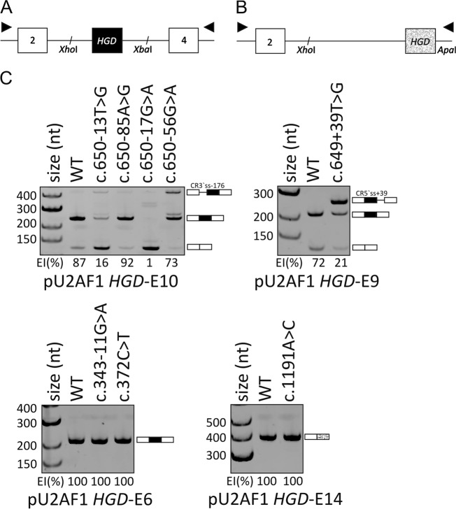
Alkaptonuria (AKU) is a rare metabolic disorder caused by a deficient enzyme in the tyrosine degradation pathway, homogentisate 1,2-dioxygenase (HGD). In 172 AKU patients from 39 countries, we identified 28 novel variants of the HGD gene, which include three larger genomic deletions within this gene discovered via self-designed multiplex ligation-dependent probe amplification (MLPA) probes. In addition, using a reporter minigene assay, we provide evidence that three of eight tested variants potentially affecting splicing cause exon skipping or cryptic splice-site activation. Extensive bioinformatics analysis of novel missense variants, and of the entire HGD monomer, confirmed mCSM as an effective computational tool for evaluating possible enzyme inactivation mechanisms. For the first time for AKU, a genotype-phenotype correlation study was performed for the three most frequent HGD variants identified in the Suitability Of Nitisinone in Alkaptonuria 2 (SONIA2) study. We found a small but statistically significant difference in urinary homogentisic acid (HGA) excretion, corrected for dietary protein intake, between variants leading to 1% or >30% residual HGD activity. There was, interestingly, no difference in serum levels or absolute urinary excretion of HGA, or clinical symptoms, indicating that protein intake is more important than differences in HGD variants for the amounts of HGA that accumulate in the body of AKU patients.


Conflict of interest statement

The authors declare that they have no conflict of interest.

Figures

Fig. 1
Fig. 1
Analysis of HGD variants with splicing reporter minigene assays in HepG2 cells. Schematic representation of (a) pU2AF1-HGD E6, pU2AF1-HGD E9, and pU2AF1-HGD E10 minigenes and (b) pU2AF1-HGD E14 minigene. XhoI/XbaI segments of HGD, containing exon 6, 9, or exon 10 (black box) and XhoI/ApaI segment of exon14 (textured box), were cloned between U2AF1 exons 2 and 4 (white boxes) or between U2AF1 exons 2 and MCS of pcDNA3.1vector, respectively. RT-PCR primers to amplify reporter transcripts are denoted by arrows. (c) Splicing pattern of wild-type and mutated HGD minigenes from panels a and b. Variants are at the top of panel; RNA products are shown schematically to the right. EI exon inclusion. c.650-17G>A shows almost a complete exon skipping, while c.650-13T>G leads to a mixture of skipped and correctly spliced products and activation of a cryptic 3′ ss-176 (CR3′ss-176). In case of c.650-56G>A variant EI is decreased to 73% and use of a cryptic 3′ ss-176 (CR3′ss-176) is more evident. c.649+39T>G creates de novo donor splice site at position +1 relative to 5′ss consensus sequence that is stronger than the authentic counterparts and causes inclusion of part of the intron 9 into the transcript
Fig. 2
Fig. 2
Noncovalent interaction network of residues involved in variants that disrupt HGD function by different mechanisms: (ab) directly affecting active site (Y350 and P332); (c, d) affecting protomer stability (W97 and G205); and (e, f) affecting protein–protein affinity and hemaxer formation (R336 and G185). Hydrogen bonds are depicted as red dashes, ionic interaction as yellow dashes, π interactions as gray disks, and other polar interactions as gray dashes. Interface residues (from a different chain) are colored in dark gray
Fig. 3
Fig. 3
Position of missense variants within HGD monomer and their conservation. (a) Position of 111 missense variants within HGD protomer shows that the 93 residues which are mutated in AKU (shown as spheres) tend to be the most highly conserved, missense variant intolerant residues (blue conserved/intolerant—red variable/tolerant). (b) The incidence of missense variants (y-axis) at the positions with different Shannon entropy rates (x-axis) divided into intervals of 0.1. Solid line (“All”) represents values calculated on the total number of positions; dotted line (“mut”) are values calculated on the 93 positions that carry one or more variants. The graph can be divided into three regions: (i) H.10 between 0 and 0.2: positions with high probability of pathogenic variants affecting function; it includes highly conserved residues, which are hit by the large part of missense variants; (ii) H.10 between 0.2 and 0.8: positions with high probability to be affected by variants compromising function and need to be analyzed in each individual case; (iii) H.10 between 0.8 and 1: predicted variants-free positions. (c) MTR (missense tolerance ratio) plot for HGD. MTR is a new scoring system describing population-based observations as opposed to homolog-based alignments. Regions in red achieved a study-wide FDR < 0.05. MTR = 1, depicted by the blue dashed line. Multiple gene-specific estimates are also depicted, including a gene’s median MTR (black dashed line), 25th percentile MTR (dark green dashed line), and 5th percentile lowest MTR estimates (orange dashed line). The majority of pathogenic genetic disease variants normally fall below the 25th percentile
Fig. 4
Fig. 4
Frequency of the variants affecting different structural regions within HGD protein: the number of the mutated positions within the specific region was divided by the number of all residues that represent given region. The overall variant incidence was calculated by dividing the total number of positions mutated (93) by total number of HGD residues (445 AA). Each structural regions is characterized by aminoacidic properties

References

    1. Garrod AE. Croonian lectures on inborn errors of metabolism, lecture II: alkaptonuria. Lancet. 1908;2:73–79.
    1. La DuBN, Zannoni VG, Laster L, Seegmiller JE. The nature of the defect in tyrosine metabolism in alcaptonuria. J Biol Chem. 1958;230:251–60. - PubMed
    1. Al-Sbou M, Mwafi N. Nine cases of Alkaptonuria in one family in southern Jordan. Rheumatol Int. 2012;32:621–5. doi: 10.1007/s00296-010-1701-1. - DOI - PubMed
    1. Janocha S, Wolz W, Srsen S, et al. The human gene for alkaptonuria (AKU) maps to chromosome 3q. Genomics. 1994;19:5–8. doi: 10.1006/geno.1994.1003. - DOI - PubMed
    1. Sakthivel S, Zatkova A, Nemethova M, Surovy M, Kadasi L, Saravanan MP. Mutation screening of the HGD gene identifies a novel alkaptonuria mutation with significant founder effect and high prevalence. Ann Hum Genet. 2014;78:155–64. doi: 10.1111/ahg.12055. - DOI - PubMed

Publication types

Substances