Lung cancer imaging methods in China from 2005 to 2014: A national, multicenter study
- PMID: 30737899
- PMCID: PMC6449240
- DOI: 10.1111/1759-7714.12988
Lung cancer imaging methods in China from 2005 to 2014: A national, multicenter study
Abstract
Background: The study was conducted to examine changes in diagnostic and staging imaging methods for lung cancer in China over a 10-year period and to determine the relationships between such changes and socioeconomic development.
Methods: This was a hospital-based, nationwide, multicenter retrospective study of primary lung cancer cases. The data were extracted from the 10-year primary lung cancer databases at eight tertiary hospitals from various geographic areas in China. The chi-squared test was used to assess the differences and the Cochran-Armitage trend test was used to estimate the trends of changes.
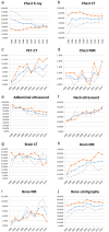
Results: A total of 7184 lung cancer cases were analyzed. Over the 10-year period, the utilization ratio of diagnostic imaging methods, such as chest computed tomography (CT) and chest magnetic resonance imaging (MRI), increased from 65.79% to 81.42% and from 0.73% to 1.96%, respectively, while the utilization ratio of chest X-ray declined from 50.15% to 30.93%. Staging imaging methods, such as positron emission tomography-CT, neck ultrasound, brain MRI, bone scintigraphy, and bone MRI increased from 0.73% to 9.29%, 22.95% to 47.92%, 8.77% to 40.71%, 42.40% to 62.22%, and 0.88% to 4.65%, respectively; abdominal ultrasound declined from 83.33% to 59.9%. These trends were more notable in less developed areas than in areas with substantial economic development.
Conclusion: Overall, chest CT was the most common radiological diagnostic method for lung cancer in China. Imaging methods for lung cancer tend to be used in a diverse, rational, and regionally balanced manner.
Keywords: China; imaging method; lung cancer; trend.
© 2019 The Authors. Thoracic Cancer published by China Lung Oncology Group and John Wiley & Sons Australia, Ltd.
Figures

 ) Nationwide, (
 ) high economic level areas, and (
 ) low high economic level areas.
References
-
- Torre LA, Siegel RL, Jemal A. Lung cancer statistics. Adv Exp Med Biol 2016; 893: 1–19. - PubMed
-
- Siegel RL, Miller KD, Jemal A. Cancer statistics, 2017. CA Cancer J Clin 2017; 67: 7–30. - PubMed
-
- Chen W, Zheng R, Baade PD et al Cancer statistics in China, 2015. CA Cancer J Clin 2016; 66: 115–32. - PubMed
-
- Jemal A, Tiwari RC, Murray T et al Cancer statistics, 2004. CA Cancer J Clin 2004; 54: 8–29. - PubMed
Publication types
MeSH terms
LinkOut - more resources
Full Text Sources
Medical

