. 2019 Apr 15:404:484-498.
doi: 10.1016/j.neuroscience.2019.01.034. Epub 2019 Feb 7.

Functionalized Phenylbenzamides Inhibit Aquaporin-4 Reducing Cerebral Edema and Improving Outcome in Two Models of CNS Injury

Affiliations

Functionalized Phenylbenzamides Inhibit Aquaporin-4 Reducing Cerebral Edema and Improving Outcome in Two Models of CNS Injury

George W Farr et al. Neuroscience. .

Abstract

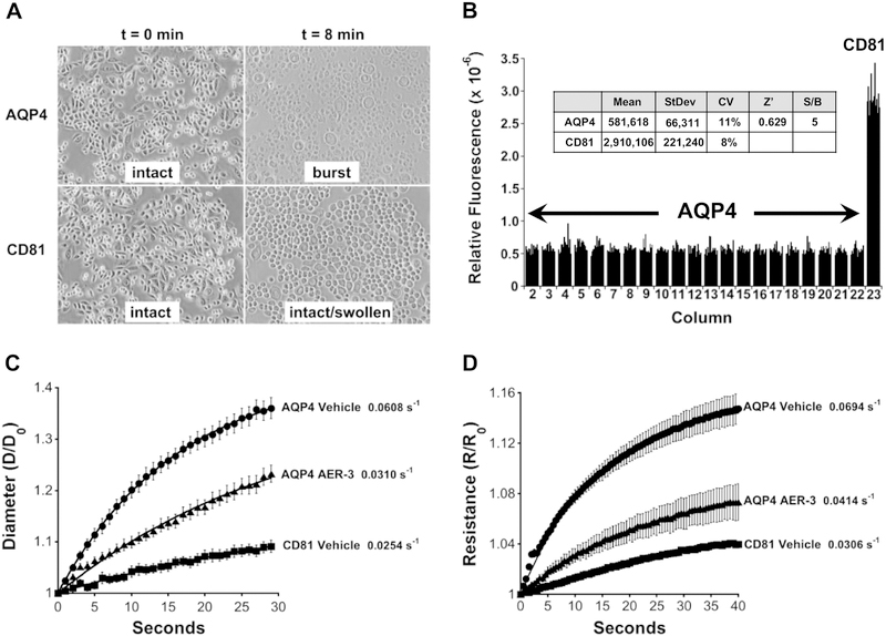
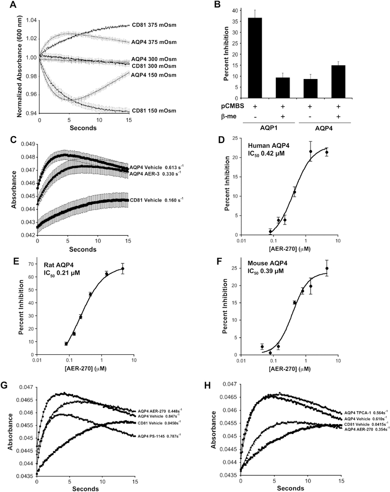
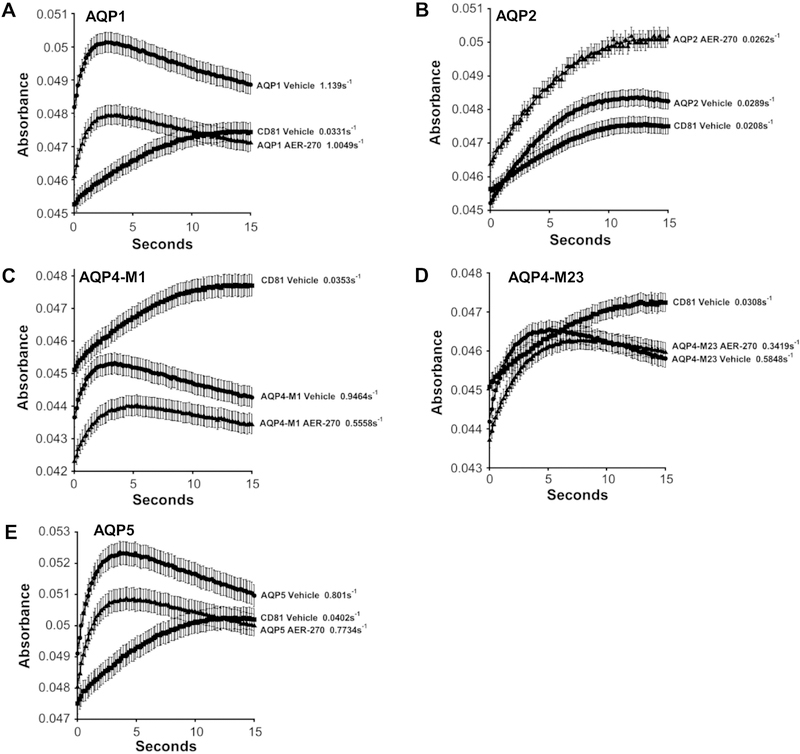
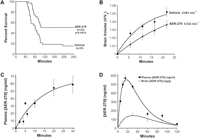
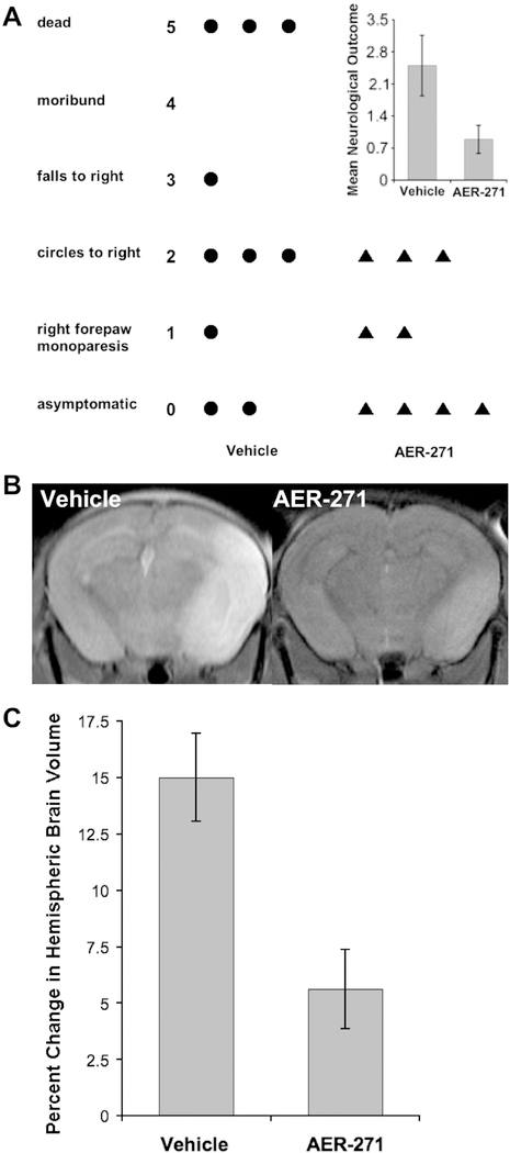
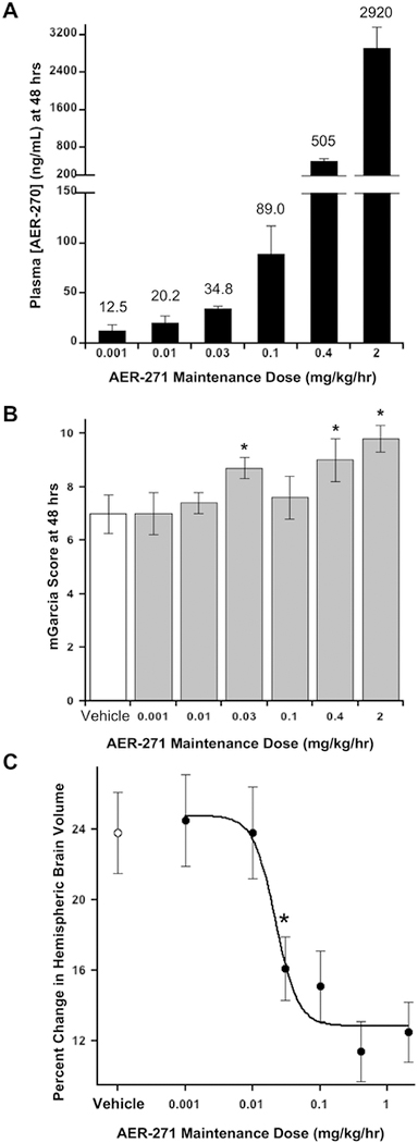
Cerebral edema in ischemic stroke can lead to increased intracranial pressure, reduced cerebral blood flow and neuronal death. Unfortunately, current therapies for cerebral edema are either ineffective or highly invasive. During the development of cytotoxic and subsequent ionic cerebral edema water enters the brain by moving across an intact blood brain barrier and through aquaporin-4 (AQP4) at astrocyte endfeet. Using AQP4-expressing cells, we screened small molecule libraries for inhibitors that reduce AQP4-mediated water permeability. Additional functional assays were used to validate AQP4 inhibition and identified a promising structural series for medicinal chemistry. These efforts improved potency and revealed a compound we designated AER-270, N-[3,5-bis (trifluoromethyl)phenyl]-5-chloro-2-hydroxybenzamide. AER-270 and a prodrug with enhanced solubility, AER-271 2-{[3,5-Bis(trifluoromethyl) phenyl]carbamoyl}-4-chlorophenyl dihydrogen phosphate, improved neurological outcome and reduced swelling in two models of CNS injury complicated by cerebral edema: water intoxication and ischemic stroke modeled by middle cerebral artery occlusion.

Keywords: AQP4 inhibitor; MCAo; cerebral edema; cytotoxic edema; ischemic stroke; water intoxication.


Conflict of interest statement

CONFLICTS OF INTEREST

G.W.F., C.H.H., S.M.F., J.M.D., A.G.A., J.M.B., P.R.M., W.F.B and M.F.P. were employees of and/or received compensation from Aeromics, Inc. All other authors declare they have no conflict of interest.

Figures

Fig. 1.
Fig. 1.
High throughput screen for inhibitors of AQP-4-mediated water permeability and cell swelling assays. (A) AQP4 imparts elevated osmotic sensitivity. CHO cells expressing human AQP4-M23 (AQP4) swell and burst within 8 min when exposed to a hypoosmotic shock (deionized water), whereas cells expressing a control membrane protein CD81 swell but remain intact. (B) High throughput assay for screening inhibitors of AQP4. The viability of cells exposed to hypoosmotic shock for 5.5 min, was measured using calcein-AM during recovery at normal osmolarity. Relative fluorescent intensity of calcein is plotted according to plate column position for CHO cells grown in a 384-well plate. AQP4 expressing cells show poor viability, lower fluorescence relative to CD81 expressing cells, and inhibitors of AQP4-mediated water permeability improve viability. Inset: 5.5 min of osmotic shock provided an optimal signal-to-noise ratio and plate statistics (Coefficient of Variation: CV = StDev/Mean; Z’ = 1 – 3[AQP4 StDev + CD81 StDev]/[AQP4 Mean - CD81 Mean]; n=14 for both AQP4 and CD81). (C) Phenylbenzamide AER-3 inhibits AQP4-mediated water movement assessed by changes in cell diameter observed with video microscopy during osmotic shock. CHO cells expressing CD81 or AQP4-M23 were given a hypoosmotic shock (deionized water), and cell diameters measured at the indicated times. Normalized mean diameters (D/Do) are plotted at the indicated times ± SEM (n=10 cells). (D) Inhibition of AQP4-mediated water movement by AER-3 assayed by Cell Volume Cytometry to measure cell volume change. CHO cells expressing AQP4-M23 or CD81 were exposed to a hypoosmotic shock and cell swelling monitored by comparing Initial Resistance (Ro) to Resistance (R) at subsequent time points using a microfluidics system. Normalized mean resistance (R/Ro) plotted ± SEM (n=4). For C and D, rate constants indicated adjacent to the fitted data.
Fig. 2.
Fig. 2.
Light scattering detects water movement in CHO cells expressing aquaporins providing an efficient assay for detecting aquaporin inhibitors. (A) AQP4 expressing CHO cells, grown as a confluent monolayer on 96-well plates, give predictable changes in light scattering in response to osmotic stress. AQP4 and CD81 expressing CHO cells were exposed to either hyperosmotic (375 mOSM), isosmotic or hypoosmotic (150 mOSM) conditions by the addition of an equal volume of HBSS adjusted to 450 mOSM using mannitol, unadjusted HBSS or water, respectively. Light scattering detected by change in absorbance at 600 nm. Mean of normalized values plotted versus time ± SEM (n=16 wells). (B) Inhibition of AQP1 but not AQP4 by mercury demonstrates utility of light scattering method for detecting inhibitors of aquaporin-based water movement. AQP1 (mercury sensitive), AQP4 (mercury insensitive) and CD81 expressing CHO cells were incubated in HBSS containing 0.3 mM p-chloromercuribenzenesulfonic acid (pCMBS) for 30 min at 37°C. Cells were subsequently incubated for 30 min in fresh HBSS with or without 5 mM β-mercaptoethanol (β-me). Changes in water permeability were then determined using Percent Inhibition calculated from light scattering data after a hyperosmotic shock, 300 to 415 mOsm, as describe in Experimental Procedures. Mean Percent Inhibition displayed ± SEM (n=8 wells). (C) Light scattering detects inhibition of AQP4-mediated water permeability by early hit AER-3. Cells were exposed to a hyperosmotic shock. Mean absorbance plotted at the indicated times ± SEM (n=8 wells). Rate constants for the rise in the curves are shown. (D-F) Lead compound AER-270 inhibits AQP4 from human, rat and mouse with similar potency. Using CHO cells expressing AQP4-M23 (AQP4) derived from human, rat (Rattus norvegicus) and mouse (Mus musculus) the IC50s of AER-270 were investigated using the light scattering assay as described in (C) above. Mean Percent Inhibition ± SEM (n=8 wells). (G-H) Kinase inhibitors selective for IKK-β do not affect AQP4-mediated water permeability. The effects of IKK-β inhibitors on AQP4-based water permeability were investigated using the light scattering assay. As a positive control 10 μM AER-270 and a Vehicle control were used to confirm AQP4 inhibition. (G) 5 μM PS-1145 and (H) 10 μM TPCA-1. Mean absorbance plotted at the indicated times ± SEM (n=8 wells).
Fig. 3.
Fig. 3.
Lead compound AER-270 preferentially inhibits AQP4. The indicated human aquaporins were stably expressed in CHO cell lines and assayed for water permeability with and without 10 μM AER-270 using the light scattering assay. (A) AQP1, (B) AQP2, (C) AQP4-M1, (D) AQP4-M23 and (E) AQP5. Mean absorbance plotted at the indicated times ± SEM (n=16 wells). Rate constants for the rise in each curve are shown. Percent Inhibition: AQP1 13.2±1.1%, AQP2 not determined, AQP4-M1 44.9±1.4%, AQP4-M23 48.8±1.6% and AQP5 4.5±0.7%.
Fig. 4.
Fig. 4.
AER-270 reduces the effects of water intoxication in mice and pharmacokinetics of AER-270 and its prodrug AER-271. (A) AER-270 improves survival from water intoxication. Mice were given an IP injection of deionized water (20% body weight): Vehicle control (0.1% DMSO) or AER-270 (0.8 mg/kg, 0.1% DMSO). General neurological changes were followed for 4 hours and time of death recorded. The percent survival is plotted as a function of time, Vehicle n=33 and AER-270 n=34 mice. P value, significance of difference between Vehicle and AER-270 curves determined by Log-Rank Test using the Kaplan-Meier estimator, p=0.0014. (B) Cerebral edema is reduced by AER-270 during water intoxication as measured by MRI brain volume analysis. T2-weighted MRI scans of mice were collected before and during water intoxication in the presence or absence of 0.8 mg/kg AER-270. Normalized mean brain volume is shown for each time point and rate constants are shown adjacent to each curve ± SEM (n=14 mice). (C) Plasma levels of AER-270 during water intoxication. The plasma concentration of AER-270 was determined by LC-MS/MS from mice injected with AER-270 in water as described in (A). Mean plasma AER-270 plotted at indicated time points ± SEM, (n=3 mice). (D) Improved solubility of prodrug AER-271 permits higher dosing and increased plasma concentrations as well as detection of AER-270 in brain tissue. Mice were given an intraperitoneal injection of 10 mg/kg AER-271 and AER-270 determined by LC-MS/MS of plasma and brain samples. Mean plasma or brain tissue concentration plotted at the indicated time points ± SEM, (n=3 mice).
Fig. 5.
Fig. 5.
Mice treated with AER-271 show improved outcomes and reduced cerebral edema in a model of ischemic stroke. (A) Improved neurological outcome following temporary, one-hour, middle cerebral artery occlusion (MCAo) in mice treated with AER-271. Mice were treated by multi-dosing (see Experimental Procedures) beginning 75 min after the occlusion was initiated using 5 mg/kg AER-271 or Vehicle and evaluated after 24 hours for general neurological outcomes (Yang et al., 1994). Inset: mean neurological outcome ± SEM, Student’s t-test P < 0.025. Vehicle n=10 mice and AER-271 n=9 mice. (B) Representative T2-weighted MR images of brains from mice receiving a temporary MCAo after 24 hrs of reperfusion. (C) Mice given a temporary MCAo and treated with AER-271 show reduced cerebral edema. Changes in hemispheric brain volume measured from T2-weighted MR images encompassing the entire brain. The mean Percent Change in Hemispheric Brain Volume (see Experimental Procedures) is displayed ± SEM for Vehicle n=7 and AER-271 n=9 mice, Student’s t-test P < 0.003.
Fig. 6.
Fig. 6.
Rats treated with AER-271 show modestly improved outcomes and reduced cerebral edema in a model of ischemic stroke. (A) Plasma AER-270 concentration after 48 hours continuous IV infusion of AER-271. An acute ischemic stroke was modelled in rats by a temporary middle cerebral artery occlusion (MCAo) for one-hour. At two-hours after initiating the injury, rats were given a loading dose administered over 30 min followed by a continuous maintenance dose of AER-271 for 48 hours by IV infusion through an external jugular vein catheter. A loading dose of 10 mg/kg was used for 2 mg/kg/hr maintenance dose; 4 mg/kg for 0.4, 0.1 and 0.03 mg/kg/hr maintenance doses; and 1 mg/kg for 0.01 and 0.001 mg/kg/hr maintenance doses. After 48 hrs of dosing plasma levels of AER-270 were quantitated by LC-MS/MS. Mean Plasma concentration of AER-270 ± SEM with mean value shown on top of each bar. AER-271 maintenance doses of 2, 0.4, 0.1, 0.03, 0.01 and 0.001 mg/kg/hr utilized n=12, n=10, n=10, n=8, n=9 and n=9 rats, respectively. (B) Improved neurological outcome after intervention with AER-271 in the rat MCAo model. After 48 hrs of reperfusion, at the indicated maintenance doses, rats were evaluated for general neurological outcomes using the modified Garcia Score (mGarcia): scale from 0 dead to 15 unaffected, assessed by multiple aspects of movement and reaction to various stimuli (Shimamura et al., 2006). Mean mGarcia ± SEM; Vehicle n=10 rats, others as indicated in (A). * Student’s t-tests P < 0.02 relative to Vehicle control. (C) Reduced cerebral edema with administration of AER-271 in rat MCAo model. Changes in hemispheric brain volume, cerebral edema, were measured from T2-weighted MR images as described in Experimental Procedures. Mean Percent Change in Hemispheric Brain Volume ± SEM displayed for the same cohorts described in (A) and (B) above. Student’s t-tests P < 0.002 for doses ≥ 0.03 mg/kg/hr relative to Vehicle control.

Similar articles

Cited by

References

    1. Agre P, King LS, Yasui M, Guggino WB, Ottersen OP, Fujiyoshi Y, Engel A, Nielsen S (2002) Aquaporin water channels--from atomic structure to clinical medicine. J Physiol (Lond) 542:3–16. - PMC - PubMed
    1. Amiry-Moghaddam M, Frydenlund DS, Ottersen OP (2004) Anchoring of aquaporin-4 in brain: molecular mechanisms and implications for the physiology and pathophysiology of water transport. Neuroscience 129:999–1010. - PubMed
    1. Anders JJ, Brightman MW (1979) Assemblies of particles in the cell membranes of developing, mature and reactive astrocytes. J Neurocytol 8:777–795. - PubMed
    1. Arac A, Blanchard V, Lee M, Steinberg GK (2009) Assessment of outcome following decompressive craniectomy for malignant middle cerebral artery infarction in patients older than 60 years of age. Neurosur Focus 26:E3. - PubMed
    1. Ayasoufi K, Kohei N, Nicosia M, Fan R, Farr GW, McGuirk PR, Pelletier MF, Fairchild RL, Valujskikh A (2018) Aquaporin 4 Blockade Improves Survival of Murine Heart Allografts Subjected to Prolonged Cold Ischemia. Am J Transplan 18:1238–1246. - PMC - PubMed

Publication types

MeSH terms