Review
. 2019 Apr;224(3):985-1008.
doi: 10.1007/s00429-019-01841-9. Epub 2019 Feb 9.

The Structural Model: a theory linking connections, plasticity, pathology, development and evolution of the cerebral cortex

Affiliations
Review

The Structural Model: a theory linking connections, plasticity, pathology, development and evolution of the cerebral cortex

Miguel Ángel García-Cabezas et al. Brain Struct Funct. 2019 Apr.

Abstract

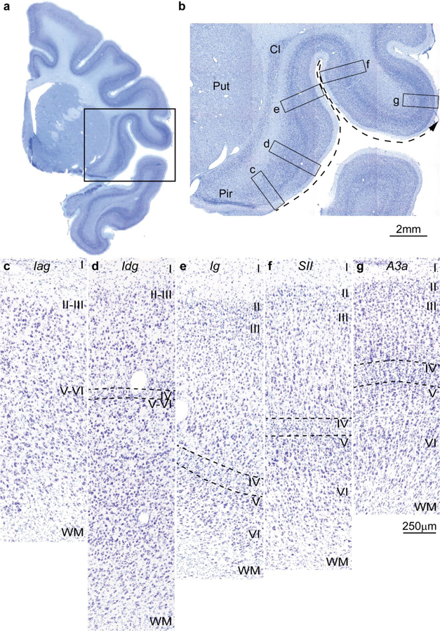
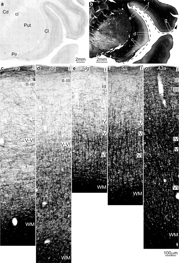
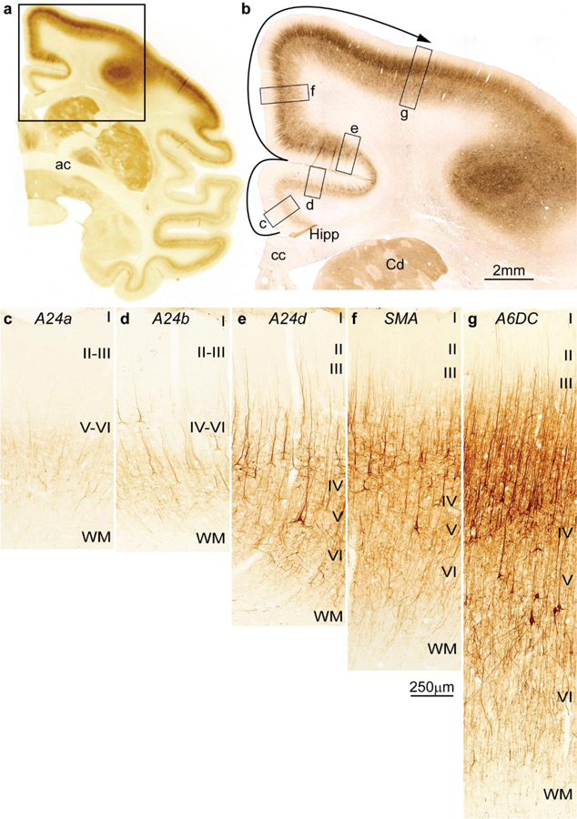
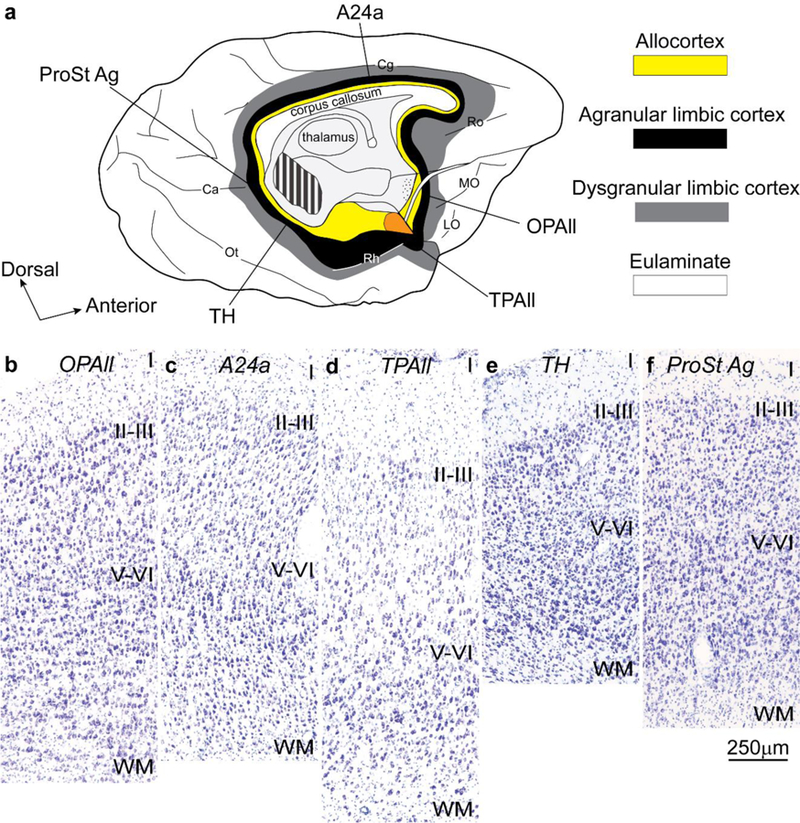
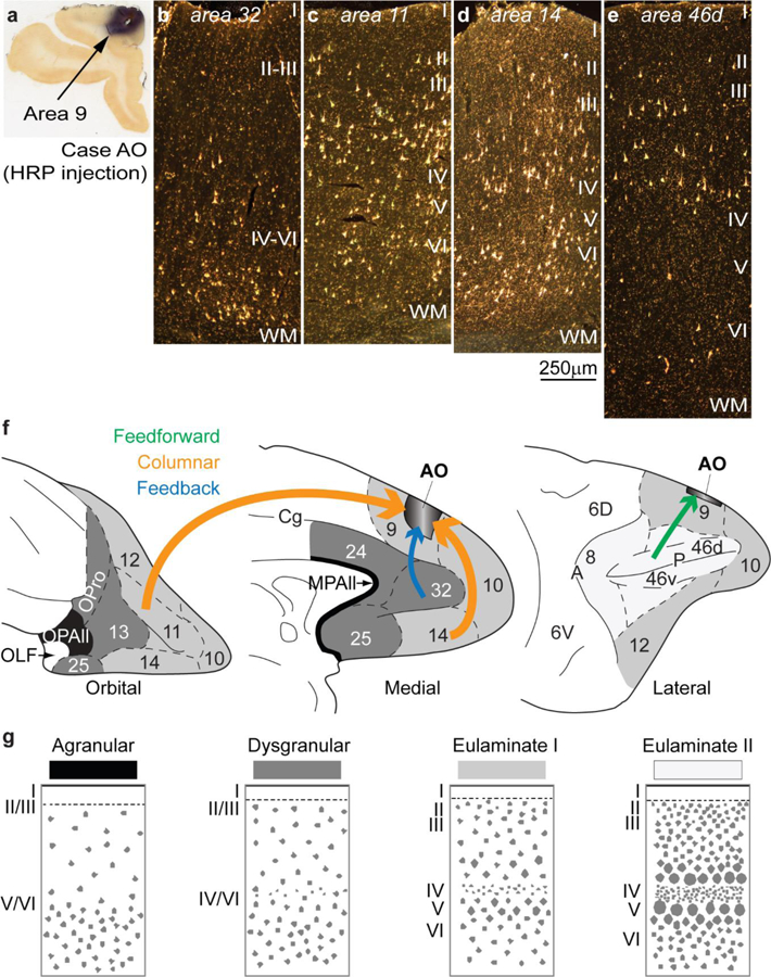
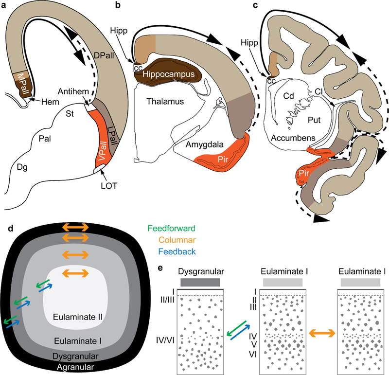
The classical theory of cortical systematic variation has been independently described in reptiles, monotremes, marsupials and placental mammals, including primates, suggesting a common bauplan in the evolution of the cortex. The Structural Model is based on the systematic variation of the cortex and is a platform for advancing testable hypotheses about cortical organization and function across species, including humans. The Structural Model captures the overall laminar structure of areas by dividing the cortical architectonic continuum into discrete categories (cortical types), which can be used to test hypotheses about cortical organization. By type, the phylogenetically ancient limbic cortices-which form a ring at the base of the cerebral hemisphere-are agranular if they lack layer IV, or dysgranular if they have an incipient granular layer IV. Beyond the dysgranular areas, eulaminate type cortices have six layers. The number and laminar elaboration of eulaminate areas differ depending on species or cortical system within a species. The construct of cortical type retains the topology of the systematic variation of the cortex and forms the basis for a predictive Structural Model, which has successfully linked cortical variation to the laminar pattern and strength of cortical connections, the continuum of plasticity and stability of areas, the regularities in the distribution of classical and novel markers, and the preferential vulnerability of limbic areas to neurodegenerative and psychiatric diseases. The origin of cortical types has been recently traced to cortical development, and helps explain the variability of diseases with an onset in ontogeny.

Keywords: Brain pathology; Cortical hierarchies; Glia; Homology; Limbic cortex; Phylogeny.


Conflict of interest statement

Disclosure of potential conflicts of interest: The authors declare that they have no conflict of interest.

Figures

Figure 1
Figure 1
Cortical types and laminar gradients of differentiation in the insula and adjacent frontal cortex of the rhesus monkey (Nissl staining). a, Coronal section of the rhesus monkey brain at the level of the frontotemporal junction stained for Nissl. b, Closer view of the insula and the frontal operculum, shows the levels of photomicrographs in c-g; the dashed arrow shows the increasing trend of laminar differentiation. c, The agranular insula (Iag) is next to the primary olfactory cortex (Pir in b) and lacks layer IV. d, Dysgranular insula (Idg) has a rudimentary layer IV (dashed lines). e, Layer IV (dashed lines) is better developed in the adjacent granular insula (Ig). f, The secondary somatosensory area (SII) has a denser layer IV (dashed lines) and better differentiation of the other layers than the granular insula. g, Area 3a, which is part of the primary somatosensory cortex, has thicker and denser layer IV (dashed lines) and its superficial layers are denser than in SII. Abbreviations: A3a: area 3a, Cl: claustrum; Iag: insula agranular; Idg: insula dysgranular; Ig: insula granular; Pir: piriform cortex in the primary olfactory cortex; Put: putamen; SII: secondary somatosensory area; WM: white matter. Roman numerals indicate cortical layers. Calibration bar in g applies to c-g.
Figure 2
Figure 2
Intracortical myelin varies systematically in parallel with laminar differentiation in the insula and adjacent frontal cortex in the rhesus monkey (Gallyas staining). a, Coronal section of the rhesus monkey brain at the level of the frontotemporal junction stained for Nissl shows the insula and the frontal operculum. b, Adjacent section to a is stained for myelin (black) and shows the levels of photomicrographs c-g; the dashed arrow shows the trend of increasing laminar differentiation. c, The agranular insula (Iag) is next to the primary olfactory cortex (Pir in a) and has comparatively fewer myelinated axons than the adjacent area. d, Dysgranular insula (Idg) is slightly better myelinated. e, The granular insula (Ig) has more myelinated axons organized into vertical bundles than Iag and Idg. f, The secondary somatosensory area (SII) has more myelinated axons than the insular areas with well-developed vertical bundles in the deep layers and abundant small horizontal axons in the middle layers. g, Area 3a (A3a) in the primary somatosensory cortex has more myelinated axons than SII in all layers. Abbreviations: A3a: area 3a, Cd: caudate; ci: internal capsule; Cl: claustrum; Iag: insula agranular; Idg: insula dysgranular; Ig: insula granular; Pir: piriform cortex in the primary olfactory cortex; Put: putamen; SII: secondary somatosensory area; WM: white matter. Roman numerals indicate cortical layers. Calibration bar in g applies to c-g.
Figure 3
Figure 3
Cortical types and laminar gradients of differentiation in the cingulate and frontal cortex of the rhesus monkey (SMI-32 staining). a, Coronal section of the rhesus monkey brain at the level of the anterior commissure (ac) stained for the nonphosphorylated neurofilament protein SMI-32. b, Higher magnification shows the cingulate and dorsal frontal cortex and the levels of photomicrographs c-g; the solid arrow shows the trend of increasing laminar differentiation. c, Agranular area 24a (A24a) in the cingulate cortex is next to the anterior extension of the hippocampal formation (Hipp in b) and has few SMI-32 labeled neurons restricted to the deep layers. d, Area 24b (A24b) in the lower bank of the cingulate sulcus has few labeled SMI-32 neurons in the deep layers and some at the bottom of layer III. e, Area 24d (A24d) in the upper bank of the cingulate sulcus has more SMI-32 neurons than the other cingulate areas in the deep layers and in layer III, demarcating a band of unstained tissue that corresponds to layer IV. f, The supplementary motor area (SMA) and (g) premotor area 6 dorsocaudal (A6DC) have more neurons labeled for SMI-32 than the cingulate areas; in both SMA and A6DC layer IV stands out as a band of unlabeled tissue between the bottom of layer III and the upper part of layer V. Abbreviations: A24a: cingulate area 24a; A24b: cingulate area 24b; A24d: cingulate area 24d; A6DC: premotor area 6 dorsocaudal; ac: anterior commissure; cc: corpus callosum; Cd: caudate; Hipp: anterior extension of the hippocampal formation; SMA: supplementary motor area; WM: white matter. Roman numerals indicate cortical layers. Calibration bar in f applies to c-g.
Figure 4
Figure 4
Neocortical areas with the simplest laminar structure form a ring at the edge (base) of the hemisphere. a, Tilted view of the macaque brain shows the medial and basal aspects of the left hemisphere. Allocortical areas (yellow) are at the edge (base) of the hemisphere; agranular limbic areas (black) are periallocortical; dysgranular limbic areas (grey) are between the periallocortex and eulaminate areas (white). Orange shows the place of the amygdala. b-f show agranular limbic areas without layer IV (Nissl staining). These areas are distributed along the periallocortical ring (black) of the neocortex at the foot of each cortical system; they have overall comparable laminar structure. Abbreviations: A24a: cingulate area 24a; Ca: calcarine fissure; Cg: cingulate sulcus; LO: lateral orbital sulcus; MO: medial orbital sulcus; OPAll: orbital periallocortex; Ot: occipitotemporal sulcus; ProSt Ag: area prostriata agranular; Rh: rhinal sulcus; Ro: rostral sulcus; TH: medial temporal area TH; TPAll: temporal periallocortex. Calibration bar in f applies to b-f.
Figure 5
Figure 5
Examples of connections predicted by the relational Structural Model. a, Injection of the neural tracer horseradish peroxidase (HRP) in area 9 of the dorsolateral prefrontal cortex in a rhesus monkey brain. b, Retrogradely labeled neurons in anterior cingulate area 32 are concentrated in the deep layers V-VI, typical of a feedback pattern of connections originating from an area with a simpler laminar structure (area 32) and directed to an area with a more elaborate laminar structure (area 9). c-d, In area 11 (c) and in area 14 (d) there are labeled neurons in superficial layers II-III and in the deep layers V-VI, typical of a columnar pattern of connections between areas of a comparable type; note absence of labeled neurons in layer IV, which do not project out of the cortex. e, Retrogradely labeled neurons in the dorsal part of area 46d are concentrated in the superficial layers II-III, typical of a predominant feedforward pattern of connections, originating from an area with a more elaborate architecture (area 46d) to an area with comparatively simpler architecture (area 9). f, Sketches of the prefrontal cortex of the rhesus monkey show the orbital (left), medial (center), and lateral (right) surfaces; agranular areas are shown in black, dysgranular areas in dark grey, and eulaminate areas are depicted progressively with lighter grey. Arrow color indicates type of connection; arrow thickness indicates relative strength of pathways; thicker arrows suggest more robust connections. g, Cortical types (agranular, dysgranular, eulaminate I, and eulaminate II) are represented in four sketches. Abbreviations: HRP: horseradish peroxidase; MPAll: medial periallocortex; OLF: primary olfactory cortex; OPAll: orbital periallocortex; OPro: orbital proisocortex. Arabic numerals correspond to prefrontal cortical areas according to Barbas and Pandya (1989). Roman numerals indicate cortical layers. Calibration bar in d applies to b-e.
Figure 6
Figure 6
Systematic variation of embryonic layers in the prospective temporal lobe of the human embryo (Nissl staining). Germinal zones and prospective cortical layers are named according to Smart et al. (2002) and Bystron et al. (2008). a-e, Photomicrographs from a human fetus of 17 weeks gestational age stained for Nissl. a, Low power photomicrograph at the level of the temporal lobe shows the levels of photomicrographs b-e. b, The germinal zones of the prospective entorhinal cortex next to the hippocampal formation are composed of thin ventricular (VZ) and subventricular (SVZ) zones; the SVZ has no sublayers. c, The SVZ of the prospective perirhinal cortex is divided into inner (ISVZ) and outer (OSVZ) zones. d, The OSVZ is more prominent in prospective lateral (eulaminate) temporal areas. e, The cell density and the thickness of the OSVZ increases progressively in a lateral direction along a line of increasing laminar elaboration. f-j, Photomicrographs from a human fetus of 20 weeks gestational age stained for Nissl. f, Low power photomicrograph at the level of the temporal lobe shows the levels of photomicrographs g-j. g, The germinal zones of the prospective entorhinal cortex next to the hippocampal formation are thicker compared to b, but the SVZ still does not show two subzones. h, The prospective perirhinal cortex has thicker VZ and SVZ; the latter is subdivided into ISVZ and OSVZ. i, Prospective lateral eulaminate areas have thicker VZ and ISVZ than prospective entorhinal and perirhinal areas; the OSVZ predominates. j, The thickness of the OSVZ increases progressively in the lateral direction consistent with increased laminar definition in the adult. Abbreviations: Cd: caudate; CP, cortical plate; ISVZ, inner subventricular zone; IZ, intermediate zone; LGE, lateral ganglionic eminence; MZ, marginal zone; OSVZ, outer subventricular zone; SP, subplate; SVZ, subventricular zone; VZ, ventricular zone. Calibration bar in i applies to b-e and g-j. [Note: This figure is a re-examination of material from an earlier paper (Reillo et al. 2011)].
Figure 7
Figure 7
Systematic variation of embryonic layers in the prospective frontal cortex of the rat embryo (Nissl staining). Germinal zones and prospective cortical layers are named according to Bystron et al. (2008) and Reillo et al. (2011). a-d, Photomicrographs from a rat embryo of 16.5 days gestational age stained for Nissl. a, Low power photomicrograph at the level of the interventricular foramen shows the levels of photomicrographs b-d. b, The germinal zones of the prospective cingulate cortex next to the hippocampal formation (Hipp in a) are composed of the ventricular (VZ), the intermediate zone (IZ) which contains many cells migrating to the cortical plate, the subplate (SP), and the cortical plate (CP) which is thin. c, d, Prospective lateral areas have slightly thicker VZ with progressive thicker CP; the IZ contains many cells migrating to the CP. At this stage, a subventricular zone (SVZ) cannot be distinguished with Nissl stain. e-h, Photomicrographs from a rat embryo of 19.5 days gestational age stained for Nissl. e, Low power photomicrograph at the level of the anterior commissure (ca) shows the levels of photomicrographs f-h. f, The prospective cingulate area shows well differentiated VZ and SVZ. g, h, In a lateral direction, the VZ and the SVZ are slightly thicker, but a clear division of the SVZ into inner and outer zones is not apparent. Abbreviations: ca: anterior commissure; cc: corpus callosum; CP, cortical plate; Hipp: anterior extension of the hippocampal formation; IZ, intermediate zone; LGE, lateral ganglionic eminence; MGE, medial ganglionic eminence; MZ, marginal zone; Pir: piriform cortex in the primary olfactory cortex; SP, subplate; SVZ, subventricular zone; VZ, ventricular zone. Calibration bar in d applies to b-d; calibration bar in h applies to f-h. [Note: This figure is an examination of material from a gift of Dr. Alan Peters].
Figure 8
Figure 8
Cortical types and laminar gradients of differentiation in the cerebral cortex of the adult rat (stained for Nissl –purple- and myelin –blue-). Areas are named according to Zilles (1985). a, b, Photomicrographs of coronal sections in an adult rat brain stained for Nissl and myelin show the levels of photomicrographs c-g (from a) and c’-g’ (from b); the solid and dashed arrows show trends of increasing laminar differentiation. c-c’, Cingulate area 2 (Cg2) and the granular retrosplenial cortex (RSG) are found next to the anterior extension of the hippocampal formation (Hipp in a, b) and lack layer IV. d-d’, Frontal cortex, area 1 (FR1, primary motor cortex) has rudimentary layer IV (dashed lines). e-e’, Parietal cortex, area 1 (Par1, primary somesthetic cortex) has identifiable layer IV (dashed lines). f-f’, The gustatory cortex (Gu) has rudimentary layer IV (dashed lines). g-g’, The posterior part of the agranular insular cortex (AIP) is next to the piriform cortex (Pir in a, b) and lacks layer IV. Abbreviations: AIP: agranular insular cortex, posterior part; cc: corpus callosum; Cd: caudate; Cg2: Cingulate area 2; FR1: Frontal cortex, area 1 (primary motor cortex); Gu: gustatory cortex; Hipp: anterior extension of the hippocampal formation; Par1: Parietal cortex, area 1 (primary somesthetic cortex); Pir: piriform cortex in the primary olfactory cortex; RSG: granular retrosplenial cortex; WM: white matter. Roman numerals indicate cortical layers. Calibration bar in b applies to a-b. Calibration bar in g’ applies to c-g and c’-g’. [Note: This figure is an examination of material from a gift of Dr. Alan Peters].
Figure 9
Figure 9
Two organizers for the expansion of the neocortex in development and evolution: the hem and antihem. a, Sketch of the mammalian telencephalon in development in coronal section shows the pallial (cortical) sectors [medial pallium (MPall), dorsal pallium (DPall), lateral pallium (LPall), and ventral pallium (VPall)], the hem, and the antihem according to the terminology of Subramanian et al. (2009), Montiel and Aboitiz (2015), and Puelles (2017) with the addition of the distinction of two parts on the MPall sector corresponding to allocortex (hippocampus) and periallocortex (agranular/dysgranular cingulate areas) in rats (b) and primates (c) based on architectonic analysis of these species in adults. The hem, found next to the roof plate, and the antihem, found in the corticostriatal junction, are secondary organizers that secrete morphogen proteins that form two overlapping gradients (solid and dashed arrows). b, Sketch of the rat adult brain in a coronal section; the adult derivatives of the developmental pallial sectors are colored as in a. The solid arrow shows the trend of laminar differentiation traced to the ancestral hippocampal cortex; the dashed arrow shows the trend of laminar differentiation traced to the ancestral olfactory cortex (Pir). c, Sketch of the adult rhesus monkey brain in coronal section; the adult derivatives of the developmental pallial sectors are tinted as in a. The solid arrow shows the dorsal trend of laminar differentiation traced to the ancestral hippocampal cortex; the dashed arrow shows the ventral trend of laminar differentiation traced to the ancestral olfactory cortex Pir). Note that DPall derivatives are more expansive than in the rat and extend to dorsal and ventral regions. d, Schematic of the primate cerebral cortex shows the arrangement of cortical types in rings. Laminar differentiation progresses from the outer or basal (black and dark grey) to the inner rings (lighter shades of grey). The edge of the cortex (black and dark grey) is actually thin compared to the greatly expanded eulaminate areas in the center. Cortical areas have stronger connections with other areas in the same ring and display columnar patterns of connections (orange arrows). Connections between areas in different rings (i.e., of different cortical type) are less strong than connections within the same ring and display feedback (blue arrows) and feedforward (green arrows) laminar patterns of connections. e, according to the Structural Model (Barbas and Rempel-Clower 1997), the laminar pattern of connections is related to the cortical type difference of the connected areas. Pathways form dysgranular to eulaminate areas are feedback (blue arrow); pathways from eulaminate to dysgranular are feedforward (green arrow); pathways between areas of comparable cortical type are columnar (orange arrow). Abbreviations: cc: corpus callosum; Cd: caudate; Cl: claustrum; Dg: Subpallial diagonal domain; DPall: dorsal pallium; Hipp: anterior extension of the hippocampal formation; LOT: lateral olfactory tract; LPall: lateral pallium; MPall: medial pallium; Pal: pallidum; Pir: piriform cortex in the primary olfactory cortex; Put: putamen; St: striatum; VPall: ventral pallium. Roman numerals indicate cortical layers.

References

    1. Abbie AA (1940) Cortical lamination in the monotremata. J Comp Neurol 72:429–467
    1. Abbie AA (1942) Cortical lamination in a polyprotodont marsupial, perameles nasuta. J Comp Neurol 76:509–536
    1. Allman J (2000) Evolving brains Scientific American Library, New York
    1. Ariëns Kappers CU, Huber GC, Crosby EC (1936) The comparative anatomy of the nervous system of vertebrates, including man Macmillan, New York,
    1. Arnold SE, Hyman BT, Flory J, Damasio AR, Van Hoesen GW (1991) The topographical and neuroanatomical distribution of neurofibrillary tangles and neuritic plaques in the cerebral cortex of patients with Alzheimer’s disease. Cereb Cortex 1:103–116 - PubMed

LinkOut - more resources