Review
. 2019:41:129-153.
doi: 10.1007/7854_2018_74.

Prefrontal Contributions to Attention and Working Memory

Affiliations
Review

Prefrontal Contributions to Attention and Working Memory

Zahra Bahmani et al. Curr Top Behav Neurosci. 2019.

Abstract

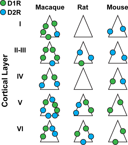
The processes of attention and working memory are conspicuously interlinked, suggesting that they may involve overlapping neural mechanisms. Working memory (WM) is the ability to maintain information in the absence of sensory input. Attention is the process by which a specific target is selected for further processing, and neural resources directed toward that target. The content of WM can be used to direct attention, and attention can in turn determine which information is encoded into WM. Here we discuss the similarities between attention and WM and the role prefrontal cortex (PFC) plays in each. First, at the theoretical level, we describe how attention and WM can both rely on models based on attractor states. Then we review the evidence for an overlap between the areas involved in both functions, especially the frontal eye field (FEF) portion of the prefrontal cortex. We also discuss similarities between the neural changes in visual areas observed during attention and WM. At the cellular level, we review the literature on the role of prefrontal DA in both attention and WM at the behavioral and neural levels. Finally, we summarize the anatomical evidence for an overlap between prefrontal mechanisms involved in attention and WM. Altogether, a summary of pharmacological, electrophysiological, behavioral, and anatomical evidence for a contribution of the FEF part of prefrontal cortex to attention and WM is provided.

Keywords: Attention; Dopamine; Working memory.


Figures

Fig. 1
Fig. 1
Changes in MT oscillatory power and spike timing during WM. (a) Schematic of the MGS task. The monkey fixates on a central fixation point (FP), and a cue stimulus appears in one of six positions arranged around the neuron’s RF location (right). The cue stimulus disappears, and the monkey maintains fixation throughout a blank delay period. Following the disappearance of the fixation point, the monkey saccades to the remembered location to receive a reward. (b) Firing rate of MT neurons does not change based on WM location. The bottom plot shows the normalized firing rate of 107 MT neurons across the course of the MGS task, when the memorized location is inside (IN, red) and outside (OUT, blue) of the neurons’ RFs. The upper-right scatter plot shows raw firing rates during the last 500 ms of the memory period, and diagonal histogram shows the distribution of firing rate changes. (c) αβ LFP power reflects WM location. The average LFP power spectrum during the memory period across frequencies (n = 480 LFP recordings), for memory IN (red) and memory OUT (blue) condition. The scatter plot shows αβ power (8–25 Hz) during memory IN vs. OUT. The diagonal histogram shows the distribution of differences in αβ power for all LFPs. (d) αβ SPL reflects WM location. The SPL for memory IN (red) and memory OUT (blue), across frequencies for all pairs of neurons and simultaneously recorded LFPs (n = 1,605 neuron-LFP pairs). Inset scatter plot shows the SPL at αβ for memory IN compared to memory OUT, with the SPL values for multiple simultaneously recorded LFPs averaged for each neuron (n = 107 neurons). Shading and error bars show standard error. Adapted from Bahmani et al. (2018)
Fig. 2
Fig. 2
Changes in MT visual sensitivity during WM. (a) During the memory period, visually evoked activity increased, but delay activity in the absence of probes was unchanged. A revised version of MGS task with probe presentation was used; visual probes appeared on 91% of trials, during both the fixation and delay periods of the MGS task. Scatter plot of WM-induced changes in the visually evoked spiking activity (probe trials) against WM-induced changes in the delay period activity (no-probe trials). Top histogram indicates the WM-induced change in delay period activity; right histogram indicates the WM-induced change in visually evoked activity. Diagonal histogram illustrates the difference between the effects of WM on firing rates in the presence and absence of visual probes. (b) RFs shift toward the WM location. Heat map showing the RF of an example MT neuron during fixation (at cross); RF of the same neuron measured while the monkey remembered a location inside of the RF, indicated by the arrow; and RF of the same neuron while the monkey remembered a location to the right of the fixation RF. Lower plots show the RF outlines of three simultaneously recorded MT neurons during fixation (left) and the delay period when the monkey remembered different locations (right). The blue outline is the RF of the neuron shown in upper plot. (c) Visual information encoded in spike phases increases near the memory location. The increase in mutual information during WM depends on the distance between the probes and the RF center or memory location. Color scale shows the change in mutual information (memory – fixation) between the spikes’ phases (αβ) and probe location for pairs of probes. The change is plotted as a function of the probes’ distance from the RF center (y-axis) and distance from the memory location (x-axis). The geometric mean of the two probe positions was used to calculate distances. Adapted from Bahmani et al. (2018) and Merrikhi et al. (2017)
Fig. 3
Fig. 3
PFC D1R manipulation enhances visual responses in V4. (a) A micro-injectrode (Noudoost and Moore 2011a) was used to deliver a small volume of D1R antagonist into the FEF. Electrical microstimulation of the FEF prior to pharmacological infusion allowed estimation of the affected area of space based on the endpoints of electrically evoked saccades (red traces). D1R antagonist infusion biased the animal’s saccades toward the RF location in a two target free-choice saccade task (right). (b) Visual responses were recorded from V4 neurons during a passive fixation task, both before and after FEF D1R manipulation (gray and red traces, respectively). Results are shown for an example V4 neuron. Following FEF manipulation, normalized responses were greater (top), orientation selectivity increased (middle), and variability decreased (bottom). (c) Summary of effects of FEF D1R manipulation for the population of V4 neurons (n = 37). In the “overlap” condition, V4 RFs corresponded with the endpoints of electrically evoked saccades. Bar graphs to the right show the change in V4 orientation selectivity, normalized response, and variability (Fano Factor) after FEF D1R manipulation compared to baseline (orange). V4 visual response magnitude and selectivity increased, while cross-trial variability decreased, following FEF D1R manipulation. Infusing GABA agonist muscimol into the FEF reduced the selectivity of V4 responses, without altering overall firing rate or variability (blue). No changes in V4 activity, selectivity, or variability were observed when the D1R manipulation occurred at an FEF site not overlapping with the V4 RFs (green) or when a D2R agonist (magenta) or saline (gray) was infused at an overlapping FEF site. In all cases D1R effects were significantly different from all other conditions. Single, double, and triple asterisks denote significance at p < 0.05, p < 0.01, and p < 0.001, respectively. Adapted from Noudoost and Moore (2011b)
Fig. 4
Fig. 4
Comparison of visual, memory, and motor activity in V4-projecting FEF neurons and the FEF population as a whole. Visual, memory, and motor selectivity were assessed using a memory-guided saccade task in which the cue appeared inside or opposite the RF of the FEF neurons being recorded (see Fig. 1a). Histograms show the distribution of average visual, memory, and motor selectivity for 1,000 ensembles of 15 FEF neurons chosen at random from the population (n = 307 non-projecting FEF neurons). Yellow arrow shows the mean selectivity for the V4-projecting FEF neurons (n = 15). Selectivity was measured based on the ROC value for trials in which the cue appeared inside vs. outside the FEF RF (during the visual, delay, or motor epochs of the task). Memory selectivity was significantly stronger in the V4-projecting FEF population, and motor selectivity was significantly weaker, compared to the non-projecting FEF population. Modified from (Merrikhi et al. 2017)
Fig. 5
Fig. 5
Distribution of D1Rs and D2Rs across cortical layers in different species. D1Rs (green) and D2Rs (blue) are more abundant in macaque than rodent species. In the macaque, D1R expression tends to decrease with cortical depth, while D2R expression increases. The opposite is true in both rodent species

References

    1. Albrecht DG (1995) Visual cortex neurons in monkey and cat: effect of contrast on the spatial and temporal phase transfer functions. Vis Neurosci 12(6):1191–1210Google Scholar - PubMed
    1. Anderson JC, Kennedy H, Martin KAC (2011) Pathways of attention: synaptic relationships of frontal eye field to V4, lateral intraparietal cortex, and area 46 in macaque monkey. J Neurosci 31(30):10872–10881. 10.1523/JNEUROSCI.0622-11.2011 - DOI - PMC - PubMed
    1. Anderson EB, Mitchell JF, Reynolds JH (2013) Attention-dependent reductions in burstiness and action-potential height in macaque area V4. Nat Neurosci 16(8):1125–1131. 10.1038/nn.3463 - DOI - PMC - PubMed
    1. Anton-Erxleben K, Carrasco M (2013) Attentional enhancement of spatial resolution: linking behavioural and neurophysiological evidence. Nat Rev Neurosci 14(3):188–200Google Scholar - PMC - PubMed
    1. Anton-Erxleben K, Stephan VM, Treue S (2009) Attention reshapes center-surround receptive field structure in macaque cortical area MT. Cereb Cortex 19(10):2466–2478. 10.1093/cercor/bhp002 - DOI - PMC - PubMed

LinkOut - more resources