. 2019 Feb 11;63(1):2998.
doi: 10.4081/ejh.2019.2998.

Morphological and ultrastructural analysis of normal, injured and osteoarthritic human knee menisci

Affiliations

Morphological and ultrastructural analysis of normal, injured and osteoarthritic human knee menisci

Michela Battistelli et al. Eur J Histochem. .

Abstract

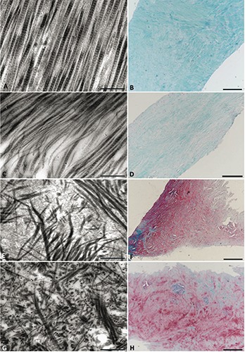
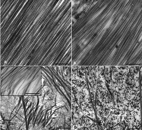
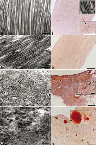
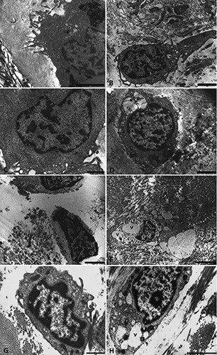
The human meniscus plays a crucial role for transmission and distribution of load across the knee, as well as shock absorption, joint stability, lubrication, and congruity. The aim of this study was to compare the complex geometry, and unique ultrastructure and tissue composition of the meniscus in healthy (control) and pathological conditions to provide understanding of structural changes that could be helpful in the future design of targetted therapies and improvement of treatment indications. We analyzed meniscus samples collected from 3 healthy multi-organ donors (median age, 66 years), 5 patients with traumatic meniscal tear (median age, 41 years) and 3 patients undergoing total knee replacement (TKR) for end-stage osteoarthritis (OA) (median age, 72 years). We evaluated the extracellular matrix (ECM) organization, the appearance and distribution of areas of calcification, and modifications of cellular organization and structure by electron microscopy and histology. The ECM structure was similar in specimens from traumatic meniscus tears compared to those from patients with late-stage OA, showing disorganization of collagen fibers and increased proteoglycan content. Cells of healthy menisci showed mainly diffuse chromatin and well preserved organelles. Both in traumatic and in OA menisci, we observed increased chromatin condensation, organelle degeneration, and cytoplasmic vacuolization, a portion of which contained markers of autophagic vacuoles. Areas of calcification were also observed in both traumatic and OA menisci, as well as apoptotic-like features that were particularly prominent in traumatic meniscal tear samples. We conclude that meniscal tissue from patients with traumatic meniscal injury demonstrate pathological alterations characteristic of tissue from older patients undergoing TKR, suggesting that they have high susceptibility to develop OA.


Conflict of interest statement

Conflict of interest: The authors report no conflicts of interest related to any aspects of the presented manuscript. The authors alone are responsible for the content and writing of the article.

Figures

Figure 1.
Figure 1.
Meniscal tissue morphology. Meniscal samples collected from one representative multi-organ donor showed collagen fibers with homogenous distribution and orientation, as observed by TEM (A) and Safranin O/Fast green staining (B). Meniscal samples collected from a 25-year-old patient during the arthroscopic procedure for meniscal tear showed collagen fibers with regular size and distribution (C) and virtually no staining for proteoglycans (D). In samples from one representative patient with meniscal tear (53 years of age) (E and F), and one representative patient with end-stage OA (G and H), TEM revealed collagen fibers with a structural disorganization of the ECM and strong staining for proteoglycans. Scale bars: A,C,E,G) 1 μm; B,F,H) 500 μm; D) 200 μm.
Figure 2.
Figure 2.
Collagen fiber organization and diameter. In samples from one representative multi-organ donor, the collagen fibers of the inner zone varied from 70 to 80 nm and showed the characteristic periodicity of collagen fibrillar organization (A). A meniscus biopsy collected from the 25-year-old patient undergoing arthroscopy for meniscal tear showed regular distribution of collagen fibers, appearing similar to that in the multiorgan donor (B). The sample from the 53-year-old patient showed meniscal degeneration with disorganized collagen fibers of between 35 to 45 nm in diameter (C and insert). The representative OA patient showed disorganized collagen fibers with diameters varying from 35 to 45 nm (D). Scale bars: 1 μm.
Figure 3.
Figure 3.
Calcium deposition. Meniscal samples collected from one representative multiorgan donor showed small calcium deposits, evaluated by Alizarin red staining (A) and rarely observed with TEM (B and insert). In meniscus samples collected from the 25-yearold patient, calcium deposits were not visible with Alizarin red (C) after trauma, but a few calcifications were detected by TEM (D). Large calcium deposits were observed in the 53-year-old patient (E and F). Extensive calcified areas were observed in one representative OA patient (G and H). Scale bars: A,C,E,G) 500 nm; B,F,H) 500 μm; D) 200 μm; insert) 50 μm.
Figure 4.
Figure 4.
Cell morphology. In the menisci from multi-organ donors, the cells embedded in the ECM were round and showed a healthy morphology, consistent with viable and metabolically active cells. In the nucleus, the chromatin appeared diffuse with small condensed areas near the nuclear membrane (A and B). In the young patient the chromatin showed small condensed areas (C and D), that were similar to those in the multi-organ donors with the cytoplasm containing small rare vacuoles (v) and autophagic vacuoles (av). In the older patients with traumatic meniscal tear meniscal cells contained nuclear chromatin that was more condensed than in the donor cells, with a specific pattern, called “chondroptosis”. In the cytoplasm abundant vacuoles were present with scarcity of cytoplasmic organelles and swollen and emptied mitochondria (E, F). Autophagic vacuoles, consistent with oxidative stress, were also present in the cytoplasm. In the representative OA patients, large areas of condensed chromatin (G) and large vacuoles within the cytoplasm were observed (H). Scale bars: A) 2 μm; B-H) 1 μm.

References

    1. Makris EA, Hadidi P, Athanasiou KA. The knee meniscus: structure-function, pathophysiology, current repair techniques, and prospects for regeneration. Biomaterials 2011;32:7411-31. - PMC - PubMed
    1. Loeser RF, Goldring SR, Scanzello CR, Goldring MB. Osteoarthritis: a disease of the joint as an organ. Arthritis Rheum 2012;64:1697-707. - PMC - PubMed
    1. Hunter DJ, Zhang YQ, Niu JB, Tu X, Amin S, Clancy M, et al. The association of meniscal pathologic changes with cartilage loss in symptomatic knee osteoarthritis. Arthritis Rheum 2006; 54:795-801. - PubMed
    1. Ding C, Martel-Pelletier J, Pelletier JP, Abram F, Raynauld JP, Cicuttini F, et al. Meniscal tear as an osteoarthritis risk factor in a largely non-osteoarthritic cohort: a cross-sectional study. J Rheumatol 2007;34:776-84. - PubMed
    1. Englund M, Roos EM, Lohmander LS. Impact of type of meniscal tear on radiographic and symptomatic knee osteoarthritis: a sixteen-year followup of meniscectomy with matched controls. Arthritis Rheum 2003;48:2178-87. - PubMed