

Review
doi: 10.1016/j.ceb.2018.12.011.
Epub 2019 Feb 8.
Recent insights into mammalian ER-PM junctions
Affiliations
- PMID: 30739879
- PMCID: PMC6462233
- DOI: 10.1016/j.ceb.2018.12.011
Item in Clipboard
Review
Recent insights into mammalian ER-PM junctions
Curr Opin Cell Biol.
2019 Apr.
Abstract
ER-PM junctions are subcellular sites where the endoplasmic reticulum (ER) and the plasma membrane (PM) are kept in close appositions, providing a platform for inter-organelle contact. These membrane contact sites are important for many physiological functions in mammalian cells, including excitation-contraction coupling, store-operated Ca2+ entry, and non-vesicular transfer of lipids between the ER and the PM. Here we review recent insights into the 3D structure and spatial organization of ER-PM junctions in mammalian cells as well as molecular mechanisms underlying the formation and functions of mammalian ER-PM junctions.
Copyright © 2019 Elsevier Ltd. All rights reserved.
Figures
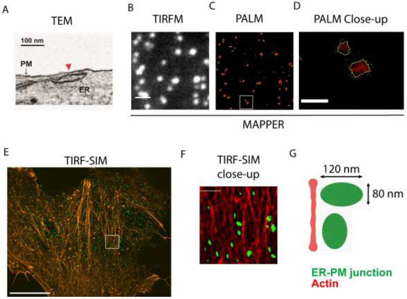
(A) A TEM image of an ER-PM junction (red arrow head) in a
MAPPER-expressing HeLa cell. (B-C) Images of the adherent surface of a
MAPPER-expressing HeLa cell acquired using TIRF microscopy (TIRFM) and PALM. (D)
A close-up from (C). (E) TIRF-SIM image of a HeLa cell with ER-PM junctions
labeled by MAPPER (green) and stained with AF568-phalloidin for F-actin
(orange). (F) A close-up from (E). (G) Diagram depicting spatial distribution of
single ER-PM junctions and cortical actin. Scale bars: 1 (B), 0.2 (D), 10 (E)
and 2 (F) μm. Figure 1A and 1B-F
were originally published in [10] and
[20].
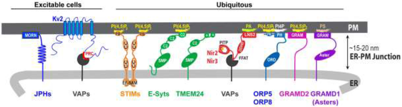
JPH, Junctophilin; MORN, membrane occupation and recognition nexus
motif; PRC, proximity restriction and clustering domain; EF-SAM, EF hand and
sterile alpha motif; CC, coiled-coil domain; PB, polybasic domain; SMP,
synaptotagmin-like mitochondrial-lipid binding protein domain; PITP,
phosphatidylinositol transfer protein domain; FFAT, two phenylalanines (FF) in
an acidic tract motif; DDHD, domain characterized by these conserved residues;
LNS2, Lipin/Ned1/Smp2 domain; ORD, OSBP-related domain; PH, pleckstrin homology
domain; GRAM, glucosyltransferases, Rab-like GTPase activators and myotubularins
domain; ASTER, START (StAR-related lipid-transfer)-like domain.
References
Publication types
MeSH terms
Substances
Grants and funding
LinkOut - more resources
Full Text Sources
Miscellaneous
