

MST1/Hippo promoter gene methylation predicts poor survival in patients with malignant pleural mesothelioma in the IFCT-GFPC-0701 MAPS Phase 3 trial
- PMID: 30739911
- PMCID: PMC6461894
- DOI: 10.1038/s41416-019-0379-8
MST1/Hippo promoter gene methylation predicts poor survival in patients with malignant pleural mesothelioma in the IFCT-GFPC-0701 MAPS Phase 3 trial
Abstract
Background: The Mesothelioma Avastin Cisplatin Pemetrexed Study (MAPS/NCT00651456) phase 3 trial demonstrated the superiority of bevacizumab plus pemetrexed-cisplatin triplet over chemotherapy alone in 448 malignant pleural mesothelioma (MPM) patients. Here, we evaluated the prognostic role of Hippo pathway gene promoter methylation.
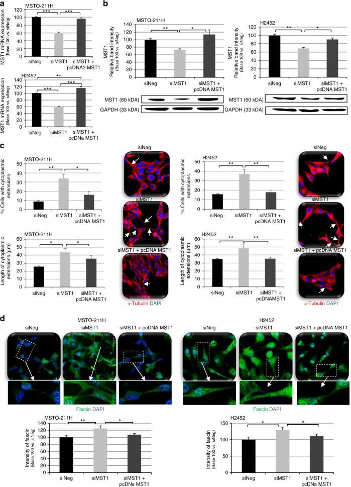
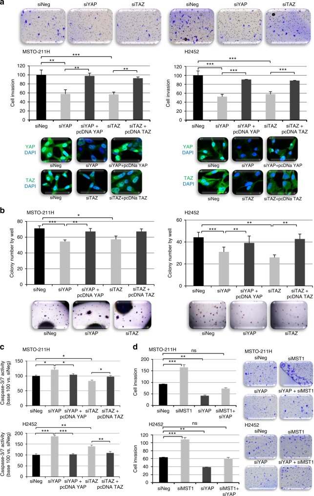
Methods: Promoter methylations were assayed using methylation-specific polymerase chain reaction in samples from 223 MAPS patients, evaluating their prognostic value for overall survival (OS) and disease-free survival in univariate and multivariate analyses. MST1 inactivation effects on invasion, soft agar growth, apoptosis, proliferation, and YAP/TAZ activation were investigated in human mesothelial cell lines.
Results: STK4 (MST1) gene promoter methylation was detected in 19/223 patients tested (8.5%), predicting poorer OS in univariate and multivariate analyses (adjusted HR: 1.78, 95% CI (1.09-2.93), p = 0.022). Internal validation by bootstrap resampling supported this prognostic impact. MST1 inactivation reduced cellular basal apoptotic activity while increasing proliferation, invasion, and soft agar or in suspension growth, resulting in nuclear YAP accumulation, yet TAZ cytoplasmic retention in mesothelial cell lines. YAP silencing decreased invasion of MST1-depleted mesothelial cell lines.
Conclusions: MST1/hippo kinase expression loss is predictive of poor prognosis in MPM patients, leading to nuclear YAP accumulation and electing YAP as a putative target for therapeutic intervention in human MPM.
Conflict of interest statement
The authors declare no competing interests.
Figures


Similar articles
-
ZIC1 is silenced and has tumor suppressor function in malignant pleural mesothelioma.J Thorac Oncol. 2013 Oct;8(10):1317-28. doi: 10.1097/JTO.0b013e3182a0840a. J Thorac Oncol. 2013. PMID: 24457242
-
Bevacizumab for newly diagnosed pleural mesothelioma in the Mesothelioma Avastin Cisplatin Pemetrexed Study (MAPS): a randomised, controlled, open-label, phase 3 trial.Lancet. 2016 Apr 2;387(10026):1405-1414. doi: 10.1016/S0140-6736(15)01238-6. Epub 2015 Dec 21. Lancet. 2016. PMID: 26719230 Clinical Trial.
-
Co-occurring Mutations of Tumor Suppressor Genes, LATS2 and NF2, in Malignant Pleural Mesothelioma.Clin Cancer Res. 2017 Jun 15;23(12):3191-3202. doi: 10.1158/1078-0432.CCR-16-1971. Epub 2016 Dec 21. Clin Cancer Res. 2017. PMID: 28003305
-
A review of bevacizumab in the treatment of malignant pleural mesothelioma.Future Oncol. 2017 Dec;13(28):2537-2546. doi: 10.2217/fon-2017-0307. Epub 2017 Sep 20. Future Oncol. 2017. PMID: 29086616 Review.
-
Prognostic and Therapeutic Implications of MicroRNA in Malignant Pleural Mesothelioma.Microrna. 2016;5(1):12-18. doi: 10.2174/2211536605666160128151018. Microrna. 2016. PMID: 26817512 Review.
Cited by
-
MST1/2 in inflammation and immunity.Cell Adh Migr. 2023 Dec;17(1):1-15. doi: 10.1080/19336918.2023.2276616. Epub 2023 Nov 1. Cell Adh Migr. 2023. PMID: 37909712 Free PMC article. Review.
-
Steps in metastasis: an updated review.Med Oncol. 2021 Jan 4;38(1):3. doi: 10.1007/s12032-020-01447-w. Med Oncol. 2021. PMID: 33394200 Review.
-
A defect of amphiregulin release predicted longer survival independently of YAP expression in patients with pleural mesothelioma in the IFCT-0701 MAPS phase 3 trial.Int J Cancer. 2022 Jun 1;150(11):1889-1904. doi: 10.1002/ijc.33997. Epub 2022 Mar 16. Int J Cancer. 2022. PMID: 35262190 Free PMC article. Clinical Trial.
-
Molecular Alterations in Malignant Pleural Mesothelioma: A Hope for Effective Treatment by Targeting YAP.Target Oncol. 2022 Jul;17(4):407-431. doi: 10.1007/s11523-022-00900-2. Epub 2022 Jul 30. Target Oncol. 2022. PMID: 35906513 Free PMC article. Review.
-
MALAT1 promotes malignant pleural mesothelioma by sponging miR-141-3p.Open Med (Wars). 2021 Nov 3;16(1):1653-1667. doi: 10.1515/med-2021-0383. eCollection 2021. Open Med (Wars). 2021. PMID: 34761116 Free PMC article.
References
Publication types
MeSH terms
Substances
LinkOut - more resources
Full Text Sources
Medical
Research Materials
Miscellaneous
