Review
. 2019 Jan 25:9:1887.
doi: 10.3389/fphys.2018.01887. eCollection 2018.

Skeletal Muscles Do Not Undergo Apoptosis During Either Atrophy or Programmed Cell Death-Revisiting the Myonuclear Domain Hypothesis

Affiliations
Review

Skeletal Muscles Do Not Undergo Apoptosis During Either Atrophy or Programmed Cell Death-Revisiting the Myonuclear Domain Hypothesis

Lawrence M Schwartz. Front Physiol. .

Abstract

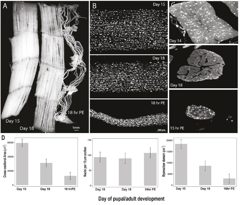
Skeletal muscles are the largest cells in the body and are one of the few syncytial ones. There is a longstanding belief that a given nucleus controls a defined volume of cytoplasm, so when a muscle grows (hypertrophy) or shrinks (atrophy), the number of myonuclei change accordingly. This phenomenon is known as the "myonuclear domain hypothesis." There is a general agreement that hypertrophy is accompanied by the addition of new nuclei from stem cells to help the muscles meet the enhanced synthetic demands of a larger cell. However, there is a considerable controversy regarding the fate of pre-existing nuclei during atrophy. Many researchers have reported that atrophy is accompanied by the dramatic loss of myonuclei via apoptosis. However, since there are many different non-muscle cell populations that reside within the tissue, these experiments cannot easily distinguish true myonuclei from those of neighboring mononuclear cells. Recently, two independent models, one from rodents and the other from insects, have demonstrated that nuclei are not lost from skeletal muscle fibers when they undergo either atrophy or programmed cell death. These and other data argue against the current interpretation of the myonuclear domain hypothesis and suggest that once a nucleus has been acquired by a muscle fiber it persists.

Keywords: Manduca sexta; autophagy; intersegmental muscle; myonuclei; sarcopenia.


Figures

Figure 1
Figure 1
Myonuclei are acquired during hypertrophy but not lost during atrophy in mouse. Micrographs of same EDL muscle fiber over time following the induction of hypertrophy (top row) and the subsequent induction of atrophy (bottom row). Fluorescently labeled oligonucleotides were used to visualize the nuclei in vivo. The dotted lines represent the sarcolemma. Scale bar = 50 μm. (Adapted from Bruusgaard et al., 2010. Used by permission of the Proceedings of the National Academy of Sciences.)
Figure 2
Figure 2
Retention of myonuclei during both atrophy and death of the intersegmental muscles (ISMs) from the moth Manduca sexta. (A) ISMs from three stages of development: homeostatic (days 15 of pupal-adult development; left); atrophic (day 18; middle); and dying (18 h post-eclosion; right). Scale bar equals ~1 mm (adapted from Schwartz et al., 2016). (B) ISMs from these same developmental stages were cleared and stained with the nuclear dye DAPI and visualized via confocal microscopy (adapted from Schwartz et al., 2016). (C) ISM fiber sections (10 μm) were stained with the nuclear dye DAPI. Note the dramatic loss of muscle protein (light gray area) during atrophy and death, but the retention of nuclei at all stages (adapted from Schwartz et al., 2016). (D) Quantification of ISM fiber volume (left), nuclear number (middle), and myonuclear domain size (right) during homeostasis, atrophy, and death. (Mean ± standard error.) (Adapted from Schwartz et al., 2016).

Similar articles

Cited by

References

    1. Aagaard P. (2004). Making muscles “stronger”: exercise, nutrition, drugs. J. Musculoskelet. Neuronal. Interact. 4, 165–174. PMID: - PubMed
    1. Abreu P., Mendes S. V., Ceccatto V. M., Hirabara S. M. (2017). Satellite cell activation induced by aerobic muscle adaptation in response to endurance exercise in humans and rodents. Life Sci. 170, 33–40. 10.1016/j.lfs.2016.11.016, PMID: - DOI - PubMed
    1. Ali S., Garcia J. M. (2014). Sarcopenia, cachexia and aging: diagnosis, mechanisms and therapeutic options—a mini-review. Gerontology 60, 294–305. 10.1159/000356760, PMID: - DOI - PMC - PubMed
    1. Alway S. E., Martyn J. K., Ouyang J., Chaudhrai A., Murlasits Z. S. (2003). Id2 expression during apoptosis and satellite cell activation in unloaded and loaded quail skeletal muscles. Am. J. Physiol. Regul. Integr. Comp. Physiol. 284, R540–R549. 10.1152/ajpregu.00550.2002, PMID: - DOI - PubMed
    1. Andrianjafiniony T., Dupre-Aucouturier S., Letexier D., Couchoux H., Desplanches D. (2010). Oxidative stress, apoptosis, and proteolysis in skeletal muscle repair after unloading. Am. J. Phys. Cell Physiol. 299, C307–C315. 10.1152/ajpcell.00069.2010, PMID: - DOI - PubMed

LinkOut - more resources