. 2019 Jan 25:10:37.
doi: 10.3389/fmicb.2019.00037. eCollection 2019.

14-3-3 Protein of Neospora caninum Modulates Host Cell Innate Immunity Through the Activation of MAPK and NF-κB Pathways

Affiliations

14-3-3 Protein of Neospora caninum Modulates Host Cell Innate Immunity Through the Activation of MAPK and NF-κB Pathways

Shan Li et al. Front Microbiol. .

Abstract

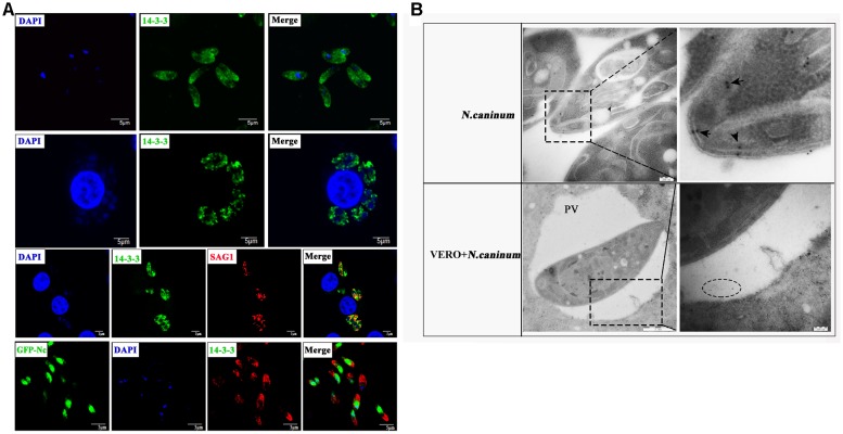
Neospora caninum is an obligate intracellular apicomplexan parasite, the etiologic agent of neosporosis, and a major cause of reproductive loss in cattle. There is still a lack of effective prevention and treatment measures. The 14-3-3 protein is a widely expressed acidic protein that spontaneously forms dimers within apicomplexan parasites. This protein has been isolated and sequenced in many parasites; however, there are few reports about the N. caninum 14-3-3 protein. Here, we successfully expressed and purified a recombinant fusion protein of Nc14-3-3 (rNc14-3-3) and prepared a polyclonal antibody. Immunofluorescence and immunogold electron microscopy studies of tachyzoites or N. caninum-infected cells suggested that 14-3-3 was localized in the cytosol and the membrane. Western blotting analysis indicated that rNc14-3-3 could be recognized by N. caninum-infected mouse sera, suggesting that 14-3-3 may be an infection-associated antigen that is involved in the host immune response. We demonstrated that rNc14-3-3 induced cytokine expression by activating the MAPK and AKT signaling pathways, and inhibitors of p38, ERK, JNK, and AKT could significantly decrease the production of IL-6, IL-12p40, and TNF-α. In addition, phosphorylated nuclear factor-κB (NF-κB/p65) was observed in wild-type peritoneal macrophages (PMs) treated with rNc14-3-3, and the protein level of NF-κB/p65 was reduced in the cytoplasm but increased correspondingly in the nucleus after 2 h of treatment. These results were also observed in deficient in TLR2-/- PMs. Taken together, our results indicated that the N. caninum 14-3-3 protein can induce effective immune responses and stimulate cytokine expression by activating the MAPK, AKT, and NF-κB signaling pathways but did not dependent TLR2, suggesting that Nc14-3-3 is a novel vaccine candidate against neosporosis.

Keywords: 14-3-3; AKT; MAPK; Neospora caninum; cytokines; immune protection.


Figures

FIGURE 1
FIGURE 1
Constructed plasmids and purified recombinant Nc14-3-3 protein. (A, Left) The Nc14-3-3 gene were amplified from cDNA using PCR. lane M: DNA Mark, lane 1: an 801-bp PCR product, arrow: an 801-bp PCR product that corresponded to Nc14-3-3. (Right) The product was cloned into the vector pGEX4T-1 and identified through restriction enzyme analysis. lane M: DNA Mark, lane 1: the pGEX-4T-1-Nc14-3-3 vector was confirmed by digestion with EcoRI and BamHI, lane 2: the pGEX-4T-1-Nc14-3-3 vector without digestion. (B) rNc14-3-3 purified using the Proteinlso GST Resin. M, protein molecular markers. Lanes 1–2, SDS-PAGE analysis of effluent and washed liquid. Lanes 3–8, the eluted buffer. (C) Western blotting analyzed of Neospora caninum lysate antigen (NLA) and rNc14-3-3 using anti-N. caninum serum (1:200). positive serum from mouse infected with N. caninum; negative serum from healthy mouse.
FIGURE 2
FIGURE 2
Immunofluorescence (A) and immunogold (B) studies of 14-3-3 protein localization. (A) Immunofluorescence microscopy of N. caninum tachyzoites or VERO cells infected with N. caninum. Scale bars: 5 μm. (B) Immunogold labeling of VERO cells infected with N. caninum tachyzoites. arrow: the gold particles within the tachyzoites, circle: the gold particles within the perivacuolar space. PV, parasitophorous vacuole.
FIGURE 3
FIGURE 3
Effect of Recombinant Nc14-3-3 on the cell viability of PMs. Mouse PMs were cultured with different protein concentrations (0, 12.5, 25, 50, 100, 200 μg/mL) for 24 h. Cell viability was determined by the CCK-8 assay. P < 0.05; ∗∗P < 0.01 for the rNc14-3-3 group versus the PBS group. CCK-8, cell counting kit.
FIGURE 4
FIGURE 4
Recombinant Nc14-3-3 activates MAPK signaling pathways in WT and TLR2-/- mouse PMs. (A) The 14-3-3 protein detected in different N. caninum-derived polypeptides fractions via Western blotting. (B) The phosphorylation levels of p38, ERK, and JNK were determined by Western blotting, the phosphorylation of p38 (at 1 h), ERK (at 1 h), and JNK (at 0.5 h) was clearly observed and the relative gray intensity analysis of the Western blotting. (C) WT and TLR2-/- mouse PMs were incubated with rNc14-3-3 protein or N. caninum lysate antigen (NLA) for 1 or 0.5 h, and the phosphorylation levels of p38 (1 h), ERK (1 h), and JNK (0.5 h) were determined by immunoblot analysis, and the relative levels of the signals from the western blot in panel. P < 0.05; ∗∗P < 0.01; ∗∗∗P < 0.001 for the rNc14-3-3 group versus the PBS group.
FIGURE 5
FIGURE 5
Recombinant Nc14-3-3-mediated cytokine production was decreased after blocking the MAPK signaling pathways. WT mouse PMs were pretreated with or without the SB203580 (p38), PD98059 (ERK) and SP600125 (JNK) inhibitors before incubated with rNc14-3-3 or N. caninum lysate antigen (NLA) for 24 h. The production of IL-6, IL-12p40, and TNF-α in the supernatants was measured by ELISA. Data are expressed as the mean ± SD from three separate experiments. P < 0.05; ∗∗P < 0.01; ∗∗∗P < 0.001 for the inhibitor-treated group versus the Nc14-3-3 group or the inhibitor-treated group versus the NLA group.
FIGURE 6
FIGURE 6
Recombinant Nc14-3-3 activates AKT signaling pathways in WT and TLR2-/- mouse PMs. (A) The phosphorylation levels of AKT in WT mouse PMs were determined by Western blotting after rNc14-3-3 or NLA treated for the indicated times. The phosphorylation of AKT was significantly increased at 1 h and the relative levels of the signals from the western blotting in panel. (B) The production of phosphorylation AKT in the WT and TLR2-/- mouse PMs was measured by Western blotting after incubated with rNc14-3-3 protein for 1 h and the relative gray intensity analysis of the Western blotting. (C) WT mouse PMs were transfected with a small-interfering RNA targeting AKT for 24 h, and AKT protein levels were significantly reduced after RNA interference, as detected by Western blotting and the relative gray intensity analysis of the Western blotting. (D) The observed changes in AKT RNA levels after siRNA interference were consistent with protein levels.Data are expressed as the mean ± SD from three separate experiments. P < 0.05; ∗∗P < 0.01; ∗∗∗P < 0.001 for the treated groups versus the untreated or PBS group.
FIGURE 7
FIGURE 7
The production of inflammatory cytokines was significantly reduced after blocking the AKT signaling pathway. (A) WT mouse PMs were pretreated with the MAP kinases inhibitors in addition or not AKT inhibitor IV before rNc14-3-3 or NLA treatment for 24 h, the production of IL-6, IL-12p40, and TNF-α in the supernatants was measured by ELISA. (B) WT mouse PMs were pretreated with AKT inhibitor IV or transfected with a small-interfering RNA targeting AKT for 24 h and then further stimulated with rNc14-3-3 for 24 h. The supernatants were removed and assayed for IL-6, IL-12p70, and TNF-α using ELISA. Data are expressed as the mean ± SD from three separate experiments. P < 0.05; ∗∗P < 0.01; ∗∗∗P < 0.001 for the inhibitor-treated group versus the Nc14-3-3 group or the inhibitor-treated group versus the NLA group.
FIGURE 8
FIGURE 8
Recombinant Nc14-3-3 induces the translocation of NF-κB/p65 subunits to the nucleus. (A, Top) WT mouse PMs were incubated with rNc14-3-3 for the indicated times and the phosphorylation levels of p65 and IκB were determined by Western blotting and relative gray intensity analysis. (Bottom) WT mouse PMs were incubated with rNc14-3-3; then, cytoplasmic and nuclear proteins were extracted separately. NF-κB/p65 protein levels were reduced in the cytoplasm but increased correspondingly in the nucleus at 2 h. (B) The effects of rNc14-3-3 or Pam3CSK4 on the translocation of NF-κB/p65 from the cytoplasm to the nucleus in the WT and TLR2-/- group were observed after incubation with rNc14-3-3 through laser scanning confocal microscopy. (C) WT and TLR2-/- mouse PMs were incubated with 50 μg/ml of the rNc14-3-3 protein for 1 h, and the phosphorylation levels of p65 and IκB were determined by Western blotting and relative gray intensity analysis.

Similar articles

Cited by

References

    1. Aitken A., Collinge D. B., van Heusden B. P., Isobe T., Roseboom P. H., Rosenfeld G., et al. (1992). 14-3-3 proteins: a highly conserved, widespread family of eukaryotic proteins. Trends Biochem. Sci. 17 498–501. 10.1016/0968-0004(92)90339-B - DOI - PubMed
    1. Almeria S., Serrano-Perez B., Darwich L., Domingo M., Mur-Novales R., Regidor-Cerrillo J., et al. (2016). Foetal death in naive heifers inoculated with Neospora caninum isolate Nc-Spain7 at 110 days of pregnancy. Exp. Parasitol. 168 62–69. 10.1016/j.exppara.2016.06.009 - DOI - PubMed
    1. Braun L., Brenier-Pinchart M. P., Yogavel M., Curt-Varesano A., Curt-Bertini R. L., Hussain T., et al. (2013). A Toxoplasma dense granule protein, GRA24, modulates the early immune response to infection by promoting a direct and sustained host p38 MAPK activation. J. Exp. Med. 210 2071–2086. 10.1084/jem.20130103 - DOI - PMC - PubMed
    1. Brokx S. J., Wernimont A. K., Dong A., Wasney G. A., Lin Y. H., Lew J., et al. (2011). Characterization of 14-3-3 proteins from cryptosporidium parvum. PLoS One 6:e14827. 10.1371/journal.pone.0014827 - DOI - PMC - PubMed
    1. Calegari-Silva T. C., Vivarini A. C., Miqueline M., Dos Santos G. R., Teixeira K. L., Saliba A. M., et al. (2015). The human parasite Leishmania amazonensis downregulates iNOS expression via NF-kappaB p50/p50 homodimer: role of the PI3K/Akt pathway. Open Biol. 5:150118. 10.1098/rsob.150118 - DOI - PMC - PubMed