

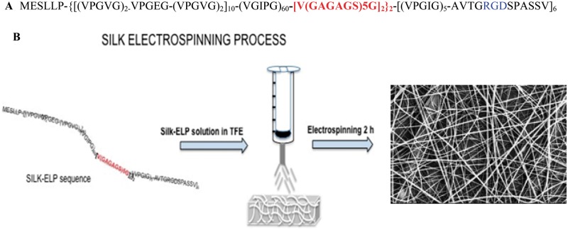
Silk-ELR co-recombinamer covered stents obtained by electrospinning
- PMID: 30740239
- PMCID: PMC6362818
- DOI: 10.1093/rb/rby022
Silk-ELR co-recombinamer covered stents obtained by electrospinning
Abstract
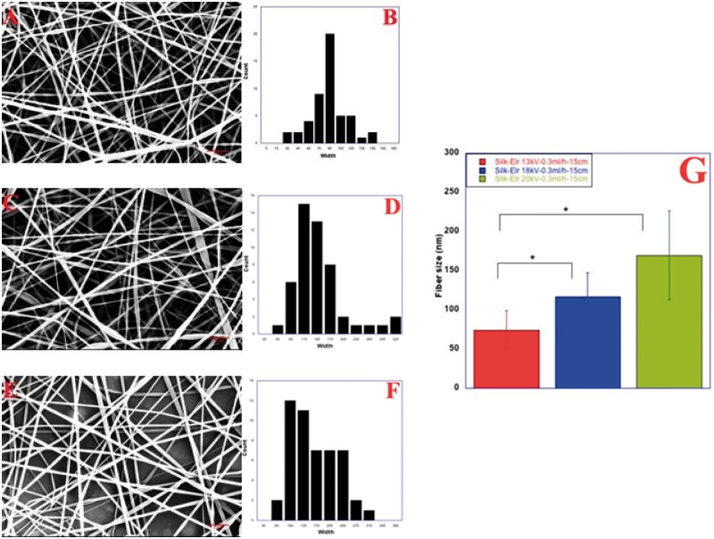
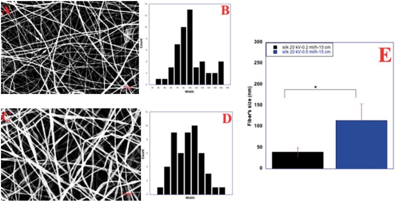
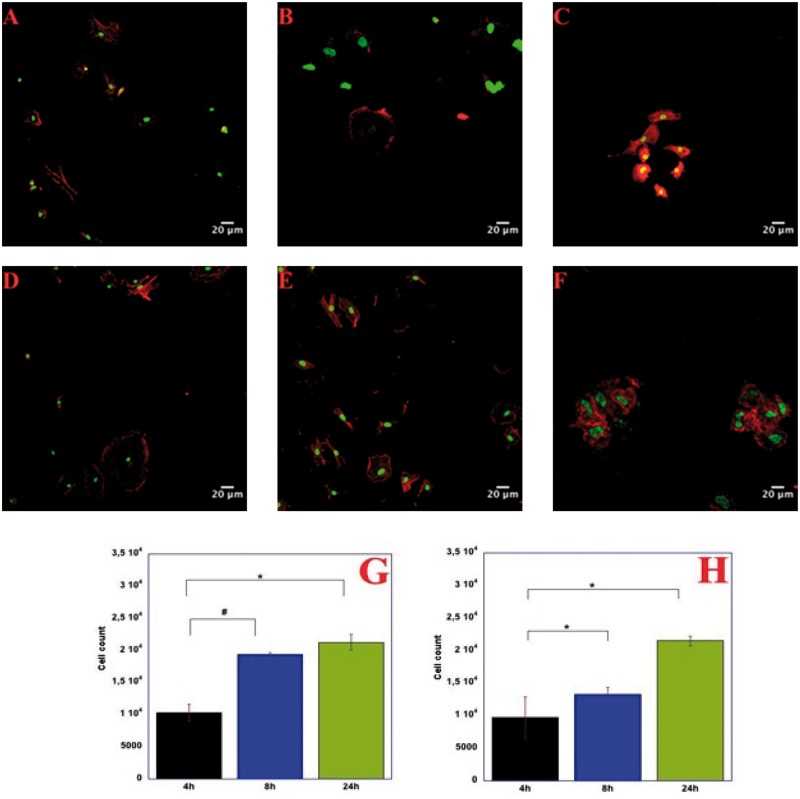
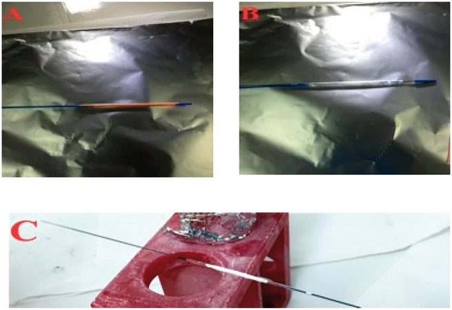
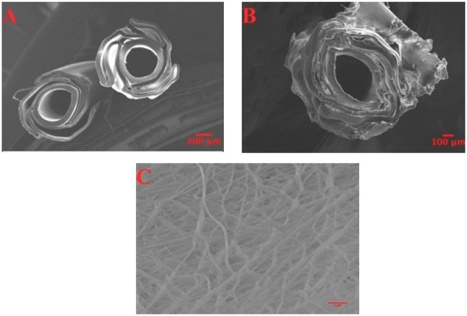
In the field of tissue engineering the choice of materials is of great importance given the possibility to use biocompatible polymers produced by means of biotechnology. A large number of synthetic and natural materials have been used to this purpose and processed into scaffolds using Electrospinning technique. Among materials that could be used for the fabrication of scaffold and degradable membranes, natural polymers such as collagen, elastin or fibroin offer the possibility to design structures strictly similar to the extracellular matrix (ECM). Biotechnology and genetic engineering made possible the advent of a new class of biopolymers called protein-based polymers. One example is represented by the silk-elastin-proteins that combine the elasticity and resilience of elastin with the high tensile strength of silk-fibroin and display engineered bioactive sequences. In this work, we use electrospinning technique to produce a fibrous scaffold made of the co-recombinamer Silk-ELR. Obtained fibres have been characterized from the morphological point of view. Homogeneity and morphology have been explored using Scanning Electron Microscopy. A thorough study regarding the influence of Voltage, flow rate and distance have been carried out to determine the appropriate parameters to obtain the fibrous mats without defects and with a good distribution of diameters. Cytocompatibility has also been in vitro tested. For the first time we use the co-recombinamer Silk-ELR for the fabrication of a 2.5 angioplasty balloon coating. This structure could be useful as a coated scaffold for the regeneration of intima layer of vessels.
Keywords: elastin-like-recombinamers; electrospinning; silk; tissue engineering.
Figures

References
-
- Alicia C, Javier AF, Matilde A et al. . Self-organized ECM-mimetic model based on an amphiphilic multiblock silk-elastin-like corecombinamer with a concomitant dual physical gelation process. Biomacromolecules 2014;15:3781–93. - PubMed
-
- Marta P, Filippo C, Valeria N et al. . Elastin-like-recombinamers multi-layered nanofibrous scaffolds for cardiovascular applications. Biofabrication 2016;8:1758–5090. - PubMed
-
- Raul M, Andrè DC, Vitor S et al. . Electrospun silk-elastin-like fibre mats for tissue engineering applications. Biomed Mater 2013;8:1748–6041. - PubMed
-
- Bini E, Knight DP, Kaplan DL. Mapping domain structures in silk from insects and spiders related to protein assembly. J Mol Biol 2004;335:27–40. - PubMed
-
- Altman GH, Diaz F, Jakuba C et al. . Silk-based biomaterials. Biomaterials 2003;24:401–16. - PubMed
LinkOut - more resources
Full Text Sources
