Review
. 2019 Jan 18;6(2):105-122.
doi: 10.15698/mic2019.02.667.

The extracellular matrix of mycobacterial biofilms: could we shorten the treatment of mycobacterial infections?

Affiliations
Review

The extracellular matrix of mycobacterial biofilms: could we shorten the treatment of mycobacterial infections?

Poushali Chakraborty et al. Microb Cell. .

Abstract

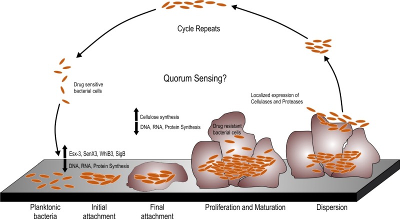
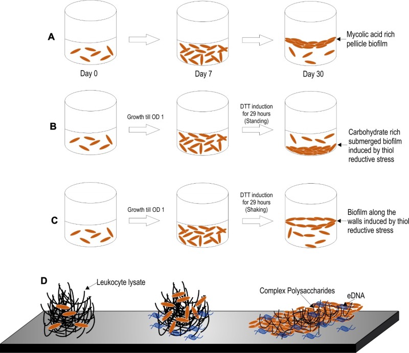
A number of non-tuberculous mycobacterium species are opportunistic pathogens and ubiquitously form biofilms. These infections are often recalcitrant to treatment and require therapy with multiple drugs for long duration. The biofilm resident bacteria also display phenotypic drug tolerance and thus it has been hypothesized that the drug unresponsiveness in vivo could be due to formation of biofilms inside the host. We have discussed the biofilms of several pathogenic non-tuberculous mycobacterium (NTM) species in context to the in vivo pathologies. Besides pathogenic NTMs, Mycobacterium smegmatis is often used as a model organism for understanding mycobacterial physiology and has been studied extensively for understanding the mycobacterial biofilms. A number of components of the mycobacterial cell wall such as glycopeptidolipids, short chain mycolic acids, monomeromycolyl diacylglycerol, etc. have been shown to play an important role in formation of pellicle biofilms. It shall be noted that these components impart a hydrophobic character to the mycobacterial cell surface that facilitates cell to cell interaction. However, these components are not necessarily the constituents of the extracellular matrix of mycobacterial biofilms. In the end, we have described the biofilms of Mycobacterium tuberculosis (Mtb), the causative agent of tuberculosis. Three models of Mtb biofilm formation have been proposed to study the factors regulating biofilm formation, the physiology of the resident bacteria, and the nature of the biomaterial that holds these bacterial masses together. These models include pellicle biofilms formed at the liquid-air interface of cultures, leukocyte lysate-induced biofilms, and thiol reductive stressinduced biofilms. All the three models offer their own advantages in the study of Mtb biofilms. Interestingly, lipids (mainly keto-mycolic acids) are proposed to be the primary component of extracellular polymeric substance (EPS) in the pellicle biofilm, whereas the leukocyte lysate-induced and thiol reductive stress-induced biofilms possess polysaccharides as the primary component of EPS. Both models also contain extracellular DNA in the EPS. Interestingly, thiol reductive stressinduced Mtb biofilms are held together by cellulose and yet unidentified structural proteins. We believe that a better understanding of the EPS of Mtb biofilms and the physiology of the resident bacteria will facilitate the development of shorter regimen for TB treatment.

Keywords: Mycobacterium; biofilms; cellulose; extracellular matrix; extracellular polymeric substance; macrocolony biofilm; pellicle biofilm; thiol reductive stress; tuberculosis pathogenesis.


Conflict of interest statement

Conflict of interest: The authors declare that they do not have any competing interests.

Figures

Figure 1
Figure 1. FIGURE 1: The biofilm formation and dispersal cycle.
The planktonic bacteria form biofilms through a series of steps, which involve the initial attachment of the cells to a substratum followed by biofilm maturation and proliferation of bacteria within the matrix and finally a part of the matured biofilm dispersing to another site for subsequent localization and attachment. During this process, bacteria undergo phenotypic changes. Several genes playing roles in virulence and redox sensing are upregulated. Biofilms formation is associated with upregulation in cellulose synthesis during maturation of the biofilms; however, localized expression of cellulases and proteases degrades the extracellular the matrix of the biofilm thereby leading to bacterial dispersal followed by start of a new cycle of biofilm formation.
Figure 2
Figure 2. FIGURE 2: Different models of biofilm formation in M. tuberculosis.
(A) Pellicle biofilm model of M. tuberculosis. The pellicle biofilm matures through several stages of development in around 5-7 weeks. These biofilms are rich in free mycolic acids. (B-C) Thiol reductive stress induced biofilm of M. tuberculosis. This model is induced by thiol reductive stress generated by reduced DTT. This polysaccharide rich biofilm of Mtb takes around 29 hours to develop. Keeping the culture flask at standing position generates a biofilm that attaches to the bottom surface of the flask (B), whereas shaking of the culture leads to biofilm formation at the liquid-air interface (C). (D) Leukocyte lysate induced biofilm model of M. tuberculosis. This eDNA rich biofilm of Mtb takes around 7 days to develop. This model may depict the biofilms formed inside the granuloma, wherein leukocyte lysate is available due to cell lysis induced by Mtb cells.
Figure 3
Figure 3. FIGURE 3: Different components of the extracellular polymeric substance of mycobacterial biofilms.
(A) The pellicle biofilm model. The inset in maroon shows the different components of the mycobacterial cell wall. The inset in pink shows the lipid based hydrophobic interactions that hold the cells in the biofilm together. The inset in blue shows the different types of mycolic acids present in the extracellular biofilm matrix, the most abundant being keto-mycolic acid. (B) The thiol reductive stress induced submerged biofilm model of Mtb. The inset in blue shows the presence of polysaccharides, cellulose macro- and micro-fibrils, different structural proteins and meshwork of other unidentified branched and unbranched polysaccharides. (C) The pellicle biofilm model of M. smegmatis and M. avium. Apart from mycolic acids, glycopeptidolipids (GPLs) play a major role in pellicle biofilm formation in these bacteria. The inset in maroon depicts the GPL structure in M. smegmatis and the same in blue depicts the GPL structure in M. avium.

References

    1. Flemming HC, Wingender J, Szewzyk U, Steinberg P, Rice SA, Kjelleberg S. Biofilms: an emergent form of bacterial life. Nat Rev Microbiol. 2016;14(9):563–575. doi: 10.1038/nrmicro.2016.94. - DOI - PubMed
    1. Hall-Stoodley L, Costerton JW, Stoodley P. Bacterial biofilms: from the natural environment to infectious diseases. Nat Rev Microbiol. 2004;2(2):95–108. doi: 10.1038/nrmicro821. - DOI - PubMed
    1. Davies D. Understanding biofilm resistance to antibacterial agents. Nat Rev Drug Discov. 2003;2(2):114–122. doi: 10.1038/nrd1008. - DOI - PubMed
    1. Parsek MR, Singh PK. Bacterial biofilms: an emerging link to disease pathogenesis. Annu Rev Microbiol. 2003;57:677–701. doi: 10.1146/annurev.micro.57.030502.090720. - DOI - PubMed
    1. Ceri H, Olson ME, Stremick C, Read RR, Morck D, Buret A. The Calgary Biofilm Device: new technology for rapid determination of antibiotic susceptibilities of bacterial biofilms. J Clin Microbiol. 1999;37(6):1771–1776. - PMC - PubMed

LinkOut - more resources