Review
. 2019 May;471(5):719-733.
doi: 10.1007/s00424-019-02260-9. Epub 2019 Feb 11.

Altered force generation and cell-to-cell contractile imbalance in hypertrophic cardiomyopathy

Affiliations
Review

Altered force generation and cell-to-cell contractile imbalance in hypertrophic cardiomyopathy

Theresia Kraft et al. Pflugers Arch. 2019 May.

Abstract

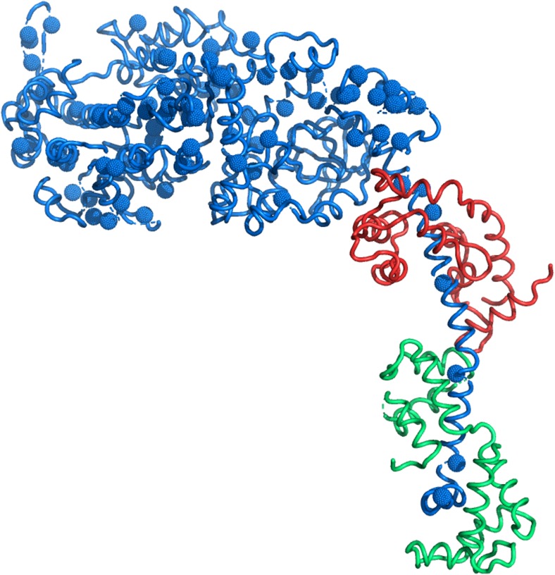
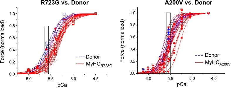
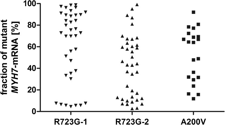
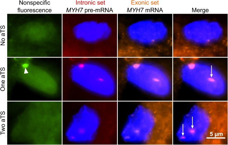
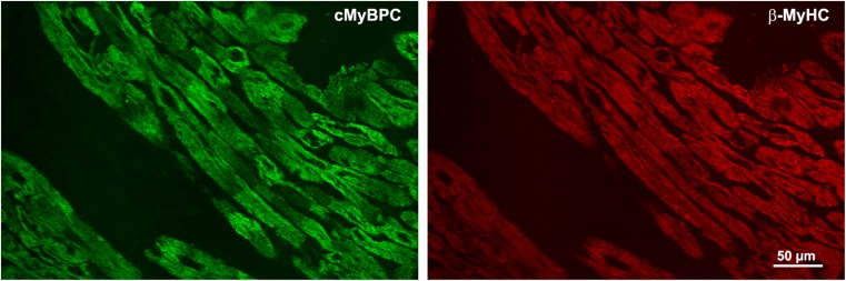
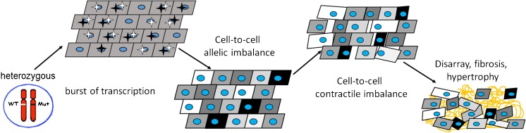
Hypertrophic cardiomyopathy (HCM) is mainly caused by mutations in sarcomeric proteins. Thirty to forty percent of identified mutations are found in the ventricular myosin heavy chain (β-MyHC). A common mechanism explaining how numerous mutations in several different proteins induce a similar HCM-phenotype is unclear. It was proposed that HCM-mutations cause hypercontractility, which for some mutations is thought to result from mutation-induced unlocking of myosin heads from a so-called super-relaxed state (SRX). The SRX was suggested to be related to the "interacting head motif," i.e., pairs of myosin heads folded back onto their S2-region. Here, we address these structural states of myosin in context of earlier work on weak binding cross-bridges. However, not all HCM-mutations cause hypercontractility and/or are involved in the interacting head motif. But most likely, all mutations alter the force generating mechanism, yet in different ways, possibly including inhibition of SRX. Such functional-hyper- and hypocontractile-changes are the basis of our previously proposed concept stating that contractile imbalance due to unequal fractions of mutated and wildtype protein among individual cardiomyocytes over time will induce cardiomyocyte disarray and fibrosis, hallmarks of HCM. Studying β-MyHC-mutations, we found substantial contractile variability from cardiomyocyte to cardiomyocyte within a patient's myocardium, much higher than in controls. This was paralleled by a similarly variable fraction of mutant MYH7-mRNA (cell-to-cell allelic imbalance), due to random, burst-like transcription, independent for mutant and wildtype MYH7-alleles. Evidence suggests that HCM-mutations in other sarcomeric proteins follow the same disease mechanism.

Keywords: Allelic imbalance; Burst-like transcription; Contractile imbalance; Hypertrophic cardiomyopathy; Weak binding states.


Conflict of interest statement

Conflict of interests

The authors declare that they have no conflict of interest.

Ethical approval

All procedures performed in studies involving human participants were in accordance with the ethical standards of the institutional and/or national research committee and with the 1964 Helsinki declaration and its later amendments or comparable ethical standards. The study was approved by the Ethics Committee of Hannover Medical School (No. 2276–2014).

Informed consent

Informed consent was obtained from all individuals included in this study.

Figures

Fig. 1
Fig. 1
HCM-mutations in the S-1 region of β-MyHC. Structure of the S-1 part of β-MyHC (blue) protein based on Rayment and colleagues [92] with mutations validated as HCM-causing by Walsh and co-workers [123]. The light chains are indicated in red (ELC) and green (RLC). Note that the mutations are distributed all over S-1
Fig. 2
Fig. 2
Contractile heterogeneity of individual cardiomyocytes from HCM-patients compared to donor cardiomyocytes. Single cardiomyocytes were isolated from frozen heart tissue of HCM-patients (red) with the mutation R723G (left) or A200V (right), respectively, and from donor individuals (blue) as controls. Cardiomyocytes were permeabilized, and after adjustment of phosphorylation levels [57, 61, 80, 114, 119], they were subjected to different calcium concentrations and the respective force generation was measured. Depicted are the forces of individual left ventricular cardiomyocytes at different calcium concentrations (force-pCa-relations), normalized to maximum force. Each symbol and curve represents a different individual cell. The boxes at physiological calcium concentration highlight the much larger variance in force generation among individual cardiomyocytes from the patients compared to controls. Figure reprinted from [82] and modified, with permission from Frontiers
Fig. 3
Fig. 3
Cell-to-cell allelic imbalance of MYH7-mRNA in three HCM patients. Individual cells were isolated from sections of cardiac tissue via laser capture microdissection. Cells were lysed and the MYH7-mRNA was amplified by single cell RT-PCR. The fractions of mutant vs. wildtype transcript were determined by densitometric analysis of allele-specific restriction digests. Depicted are the fractions of mutant MYH7-mRNA in individual cardiomyocytes from three different HCM-patients (R723G-1, R723G-2, and A200V). Each dot represents one cardiomyocyte
Fig. 4
Fig. 4
MYH7 active transcription sites in individual cardiomyocytes of an HCM-patient. Cryo-sections of 16-μm thickness were obtained from cardiac tissue of an HCM patient with the mutation R723G. Fluorescence in situ hybridization (FISH) was performed using an intronic probe set binding the pre-mRNA and an exonic probe set binding the processed mRNA. Co-localization of both fluorescently labeled probe sets in nuclei indicates active transcription sites (aTS). Shown is a cardiomyocyte without aTS (upper panel), a cardiomyocyte with one aTS (middle panel, arrow) and a cardiomyocyte with two aTS (lower panel, arrows). Note that the second signal in the middle panel (arrow head) originates from nonspecific fluorescence (left panel). Figure reprinted from [82] and modified, with permission from Frontiers
Fig. 5
Fig. 5
Heterogeneous distribution of cMyBP-C in myocardium of an HCM patient with a truncation mutation. Cryo-sections from cardiac tissue of an HCM-patient with the cMyBP-C-mutation c.927-2A>G were stained with an N-terminus-specific antibody for cMyBP-C (left panel, green) to detect the inter- and intracellular distribution of cMyBP-C. The sections were co-stained with a β-MyHC-specific antibody (right panel, red) to visualize the overall sarcomere fluorescence in the cardiomyocytes. Note the uneven distribution of cMyBP-C between and also within individual cardiomyocytes while the β-MyHC stain is much more regular
Fig. 6
Fig. 6
Contractile imbalance hypothesis for MYH7-mutations. In heterozygous HCM-patients, both MYH7 alleles are expressed burst-like; they are switched on and off in an independent and stochastic manner (active mutant and wildtype alleles are indicated by black and white stars). In adult human myocardium, in 27% of nuclei, both alleles were found switched off (no stars, i.e., no active transcription sites in scheme) [82]. Burst-like expression leads to heterogeneous fractions of wildtype and mutant mRNA in neighboring cells (indicated by differently shaded cells). This cell-to-cell allelic mRNA imbalance translates into highly heterogeneous fractions of wildtype and mutant β-MyHC protein among the cells. Due to the effect of the mutations on β-MyHC biomechanical function, the heterogeneous fractions cause imbalance in force generation from cell to cell that disrupts the cardiac syncytium over time. Stronger cells will overstretch weaker cells. This will most likely induce myocyte disarray, fibrosis, and hypertrophy

References

    1. Alamo L, Ware JS, Pinto A, Gillilan RE, Seidman JG, Seidman CE, Padron R (2017) Effects of myosin variants on interacting-heads motif explain distinct hypertrophic and dilated cardiomyopathy phenotypes. Elife 6. 10.7554/eLife.24634 - PMC - PubMed
    1. Aldag-Niebling D, Radocaj A, Hilfigker-Kleiner D, Dos Remedios C, Brenner B, Kraft T. Hypertrophic cardiomyopathy: variable expression of myosin-binding protein C from cell-to-cell and functional imbalance among individual cardiomyocytes. Biophys J. 2018;114:312A.
    1. Al-Khayat HA, Kensler RW, Squire JM, Marston SB, Morris EP. Atomic model of the human cardiac muscle myosin filament. Proc Natl Acad Sci U S A. 2013;110:318–323. - PMC - PubMed
    1. Amin AS, Giudicessi JR, Tijsen AJ, Spanjaart AM, Reckman YJ, Klemens CA, Tanck MW, Kapplinger JD, Hofman N, Sinner MF, Muller M, Wijnen WJ, Tan HL, Bezzina CR, Creemers EE, Wilde AA, Ackerman MJ, Pinto YM. Variants in the 3′ untranslated region of the KCNQ1-encoded Kv7.1 potassium channel modify disease severity in patients with type 1 long QT syndrome in an allele-specific manner. Eur Heart J. 2012;33:714–723. - PMC - PubMed
    1. Anderson RL, Trivedi DV, Sarkar SS, Henze M, Ma W, Gong H, Rogers CS, Gorham JM, Wong FL, Morck MM, Seidman JG, Ruppel KM, Irving TC, Cooke R, Green EM, Spudich JA. Deciphering the super relaxed state of human beta-cardiac myosin and the mode of action of mavacamten from myosin molecules to muscle fibers. Proc Natl Acad Sci U S A. 2018;115:E8143–E8152. - PMC - PubMed

Publication types

Substances

LinkOut - more resources