. 2019 May;234(5):700-708.
doi: 10.1111/joa.12955. Epub 2019 Feb 10.

Initial stages of development of the submandibular gland (human embryos at 5.5-8 weeks of development)

Affiliations

Initial stages of development of the submandibular gland (human embryos at 5.5-8 weeks of development)

Luis Quirós-Terrón et al. J Anat. 2019 May.

Abstract

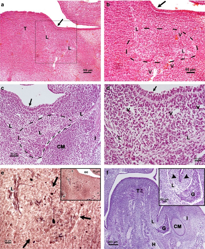
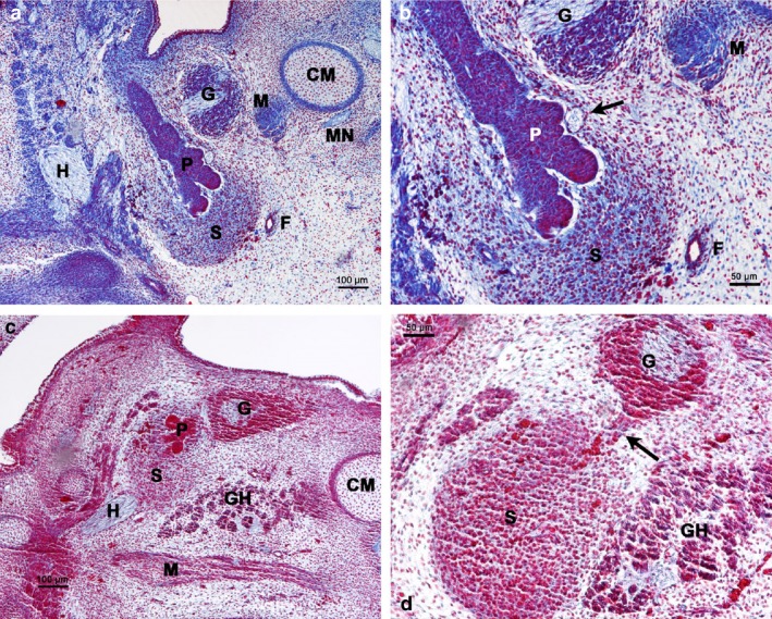
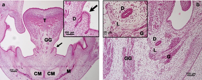
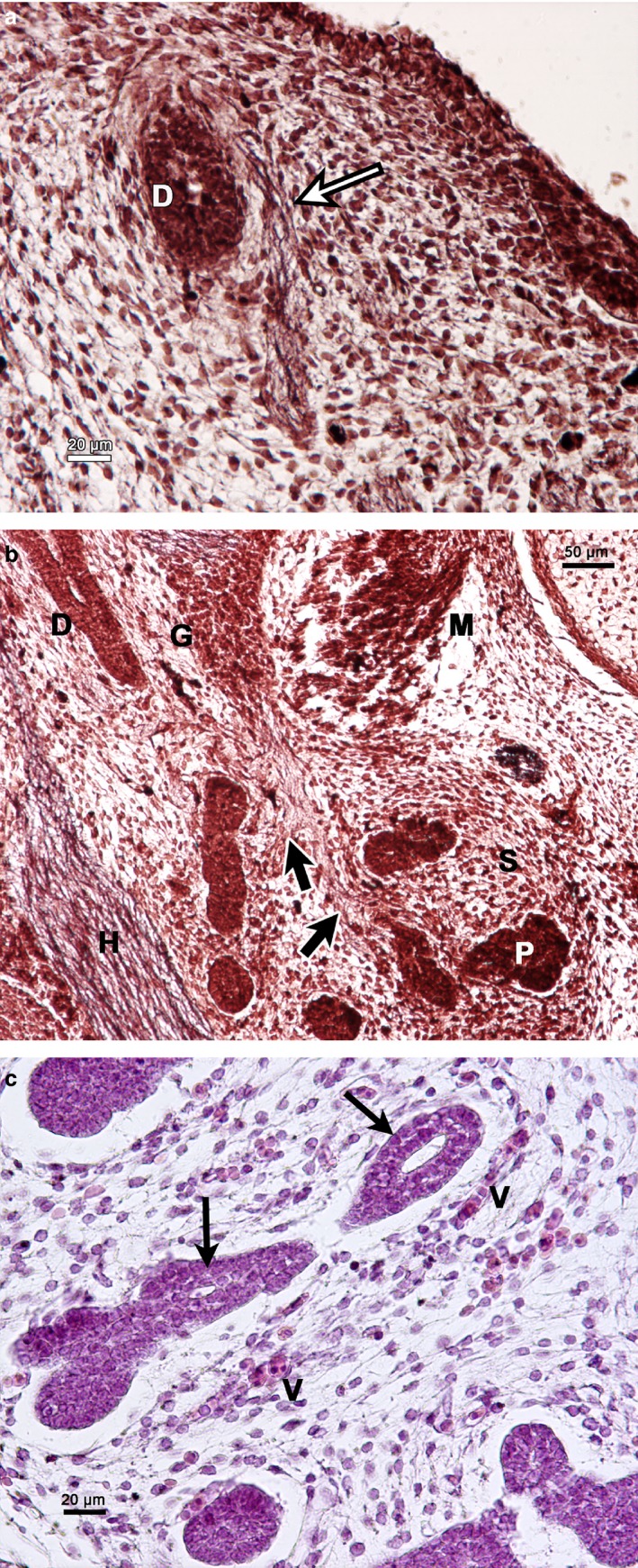
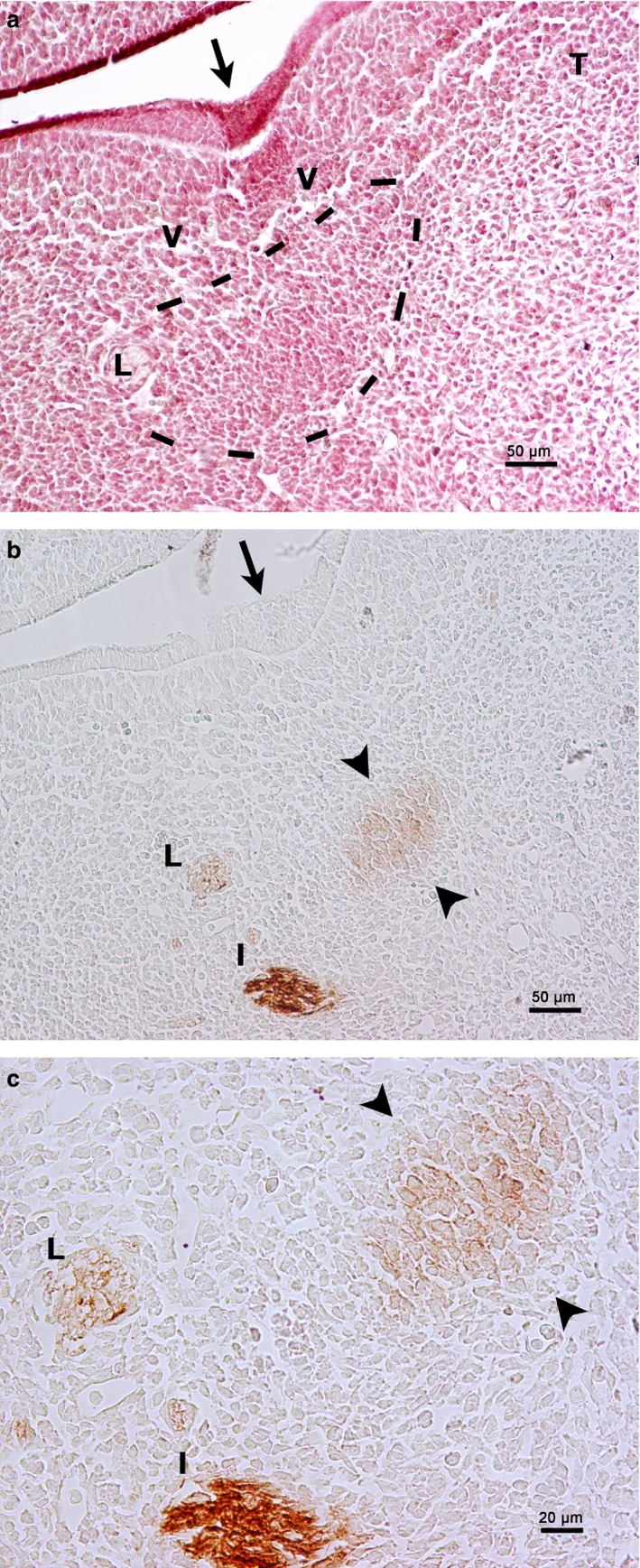
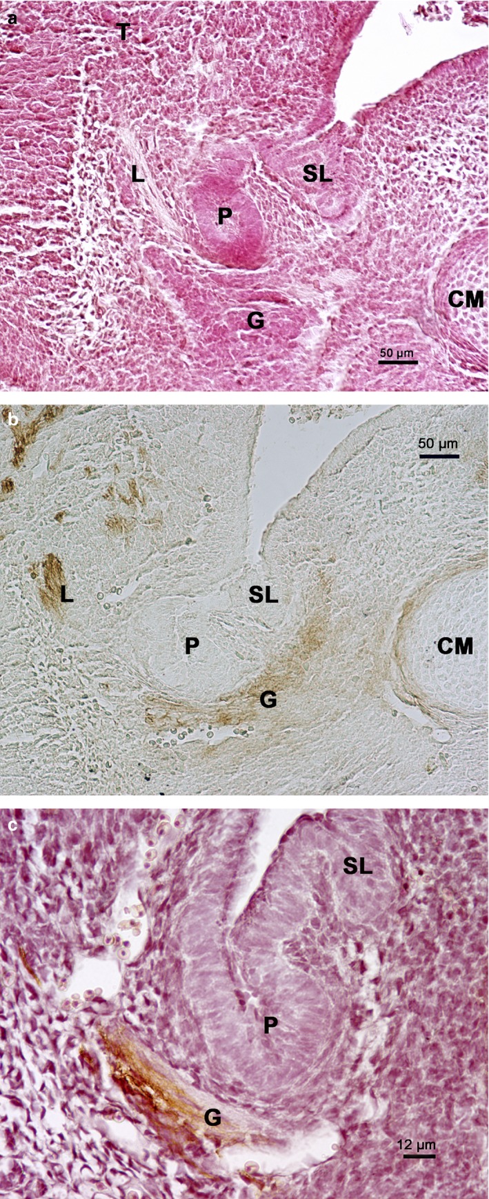
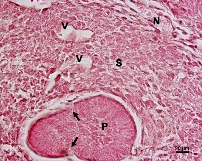
The aim of this study was to determine the main stages of submandibular salivary gland development during the embryonic period in humans. In addition, we studied submandibular salivary gland development in rats on embryonic days 14-16 and expression in the submandibular salivary gland region with the monoclonal antibody HNK-1. Serial sections from 25 human embryos with a greatest length ranging from 10 to 31 mm (Carnegie stages 16-23; weeks 5.5-8 of development) and Wistar rats of embryonic days (E) 14-16 were analysed with light microscopy. Five stages of submandibular salivary gland development were identified. The prospective stage (1), between weeks 5.5 and early week 6, is characterized by a thickening of the epithelium of the medial paralingual groove in the floor of the mouth corresponding to the primordium of the submandibular salivary gland parenchyma. At this stage, the primordium of the parasympathetic ganglion lies below the lingual nerve. The primordium of the submandibular salivary gland parenchyma is observed in rats on E14 in the medial paralingual groove with mesenchymal cells, underlying the lingual nerve. These cells are HNK-1-positive, corresponding to the primordium of the parasympathetic ganglion. The bud stage (2), at the end of week 6 in humans and on E15 in rats, is characterized by the proliferation and invagination of the epithelial condensation, surrounded by an important condensation of the mesenchyme. The pseudoglandular stage (3) at week 6.5 is characterized by the beginning of the formation of lobes in the condensed mesenchyme. The canalicular stage (4), between week 7 and 7.5, is characterized by the appearance of a lumen in the proximal part of the submandibular duct. The innervation stage (5) occurs during week 8, with the innervation of the submandibular and interlobular ducts. Nervous branches arriving from the parasympathetic ganglion innervate the glandular parenchyma. Numerous blood vessels are observed nearby. Our results suggest that submandibular salivary gland development requires interactions among epithelium, mesenchyme, parasympathetic ganglion and blood vessels.

Keywords: development; embryology; embryonic stage; submandibular salivary gland.


Conflict of interest statement

The authors have no conflict of interest to declare.

Figures

Figure 1
Figure 1
Carnegie stages 16–17. Frontal sections of the floor of the mouth. (a) Human embryo 11 mm GL. Haematoxylin‐eosin staining. The medial paralingual groove (arrow). L, lingual nerve; T, tongue. (b) Magnification of the squared area of (a). Primordium of the PSG (framed by a dashed line) in relationship to the lingual nerve (L); arrow, medial paralingual groove; V, vessel. (c). Human embryo 12 mm GL. Azan staining. The cells of the prospective area of the SMG show apical polarization (arrow). Primordium of Meckel's cartilage (CM) between the alveolar inferior nerve (I) and lingual nerve (L). Primordium of the PSG in relation to the lingual nerve (framed by a dashed line). (d). Magnification of (c). Prospective area of the SMG parenchyma (arrow). L, lingual nerve; V, vessel. (e) Human embryo 12 mm GL. Bielschowsky's staining. Primordium of the PSG (arrows) in relation to the lingual nerve (L). (e) Inset corresponding to a panoramic view. H, hypoglossal nerve; L, lingual nerve; T. tongue; OC, oral cavity. (f) Human embryo 13 mm GL. Azan staining. (f) Inset corresponding to invagination of the prospective area of the SMG parenchyma (arrowheads). CM, Meckel's cartilage; G, parasympathetic submandibular ganglion; H, hypoglossal nerve; I, inferior alveolar nerve; L, lingual nerve; T. tongue.
Figure 2
Figure 2
Carnegie stages 18–19. (a) Human embryo 16 mm GL. Frontal section of the submandibular gland region. Azan staining. Primordium of the glandular stroma (S) and glandular parenchyma (P). CM, Meckel's cartilage; F, facial artery; G, parasympathetic submandibular gland; H, hypoglossal nerve; M, mylohyoid muscle; MN, mylohyoid nerve. (b) Magnification of (a). Arrow indicates nerve. F, facial artery; G, parasympathetic submandibular ganglion; M, mylohyoid muscle; P, primordium of the glandular parenchyma; S, primordium of the glandular stroma. (c) Human embryo 15 mm GL. Sagittal section of the submandibular gland region. Haematoxylin‐eosin staining. The primordium of the parenchyma (P) and stroma (S) of the SMG is located dorsal to parasympathetic submandibular ganglion (G). CM, Meckel's cartilage; GH, geniohyoid muscle; H, hypoglossal nerve; M, mylohyoid muscle. (d) Magnification of (c). Nervous branch (arrow) from the parasympathetic submandibular ganglion (G) that reaches the primordium of the glandular stroma (S). GH, geniohyoid muscle.
Figure 3
Figure 3
Carnegie stages 20–21. Frontal section of the symphyseal region. Haematoxylin‐eosin staining. (a) Human embryo 20 mm GL. Sublingual papilla (arrow). CM, Meckel's cartilage; GG, genioglossus muscle; M, mandible; T, tongue. (a) Inset corresponds to sublingual papilla (arrow). D, Submandibular duct. (b) Human embryo 20 mm GL. (b) Inset corresponds to submandibular duct (D). G, parasympathetic submandibular ganglion; GG, genioglossus muscle; L, lingual nerve.
Figure 4
Figure 4
Carnegie stage 22–23. (a) Human embryo 26 mm GL. Bielschowsky's staining. Frontal section of the innervation (arrow) of the submandibular duct (D). (b) Human embryo 26 mm GL. Bielschowsky's staining. Frontal section of the innervation (arrows) of the glandular parenchyma (P). D, submandibular duct; G, parasympathetic submandibular ganglion; H, hypoglossal nerve; M, mylohyoid muscle; S, primordium of the glandular stroma. (c) Human embryo 31 mm GL. Azan staining. Interlobular duct with lumen (arrows). V, vessels.
Figure 5
Figure 5
Rat embryo E14. Frontal section of the floor of the mouth. (a) Haematoxylin‐eosin staining. The medial paralingual groove is beginning to form (arrow). Primordium of the PSG (framed by a dashed line) in relationship to the lingual nerve (L). T, tongue; V, vessel. (b) HNK‐1 immunoperoxidase labelling. HNK‐1 is positive to the lingual nerve (L), the inferior alveolar nerve (I) and the mesenchyme related to the lingual nerve (arrowheads). Arrow, medial paralingual groove. (c) Magnification of (a). HNK‐1 is positive to the lingual nerve (L), the inferior alveolar nerve (I) and the mesenchyme related to the lingual nerve, which corresponds to the primordium of the parasympathetic submandibular ganglion (arrowheads).
Figure 6
Figure 6
Rat embryo E15. Frontal section of the submandibular gland region. (a) Haematoxylin‐eosin staining. Primordium of the submandibular gland parenchyma (P) and sublingual gland parenchyma (SL). CM, Meckel's cartilage; G, parasympathetic submandibular ganglion; L, lingual nerve; T, tongue. (b) HNK‐1 immunoperoxidase labelling. Primordium of the submandibular gland parenchyma (P) and sublingual gland parenchyma (SL). CM, Meckel's cartilage; G, parasympathetic submandibular ganglion; L, lingual nerve. (c) HNK‐1 immunoperoxidase labelling contrasted with haematoxylin‐eosin. Primordium of the submandibular gland parenchyma (P) and sublingual gland parenchyma (SL). G, parasympathetic submandibular ganglion.
Figure 7
Figure 7
(a) Rat embryo E16. Frontal section of the primordium of the submandibular gland. Haematoxylin‐eosin staining. Lobulation begins in the primordium of the glandular parenchyma (P) (arrows). In the primordium of the glandular stroma (S), there are vessels (V) and nerves (N).

References

    1. Adameyko I, Fried K (2016) The nervous system orchestrates and integrates craniofacial development: a review. Front Physiol 7, 49. - PMC - PubMed
    1. Anatomica Terminologia (2011) Federative Committee on Anatomical Terminology. 2nd edn Stuttgart: Georg Thieme Verlag.
    1. Chi JG (1996) Prenatal development of human major salivary glands. Histological and immunohistochemical characteristics with reference to adult and neoplastic salivary glands. J Korean Med Sci 11, 203–216. - PMC - PubMed
    1. De la Cuadra‐Blanco C, Peces‐Peña MD, Mérida‐Velasco JR (2003) Morphogenesis of the human lacrimal gland. J Anat 203, 531–536. - PMC - PubMed
    1. Denny PC, Ball WD, Redman RS (1997) Salivary glands: a paradigm for diversity of gland development. Crit Rev Oral Biol Med 8, 51–75. - PubMed

LinkOut - more resources