. 2019 May;40(5):619-630.
doi: 10.1002/humu.23720. Epub 2019 Feb 28.

Cerebral hypomyelination associated with biallelic variants of FIG4

Affiliations

Cerebral hypomyelination associated with biallelic variants of FIG4

Guy M Lenk et al. Hum Mutat. 2019 May.

Abstract

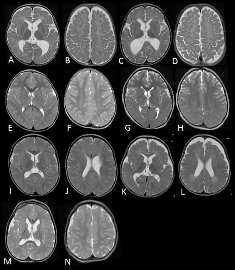
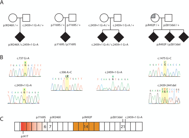
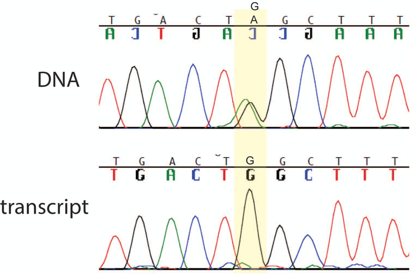
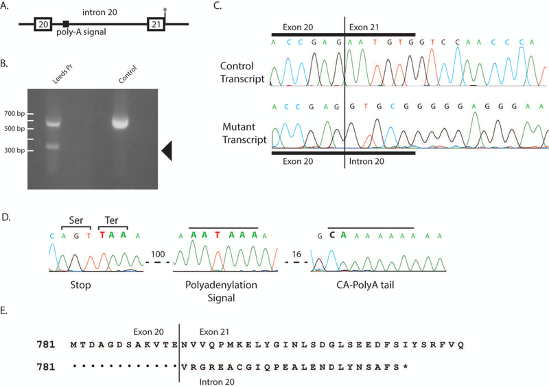
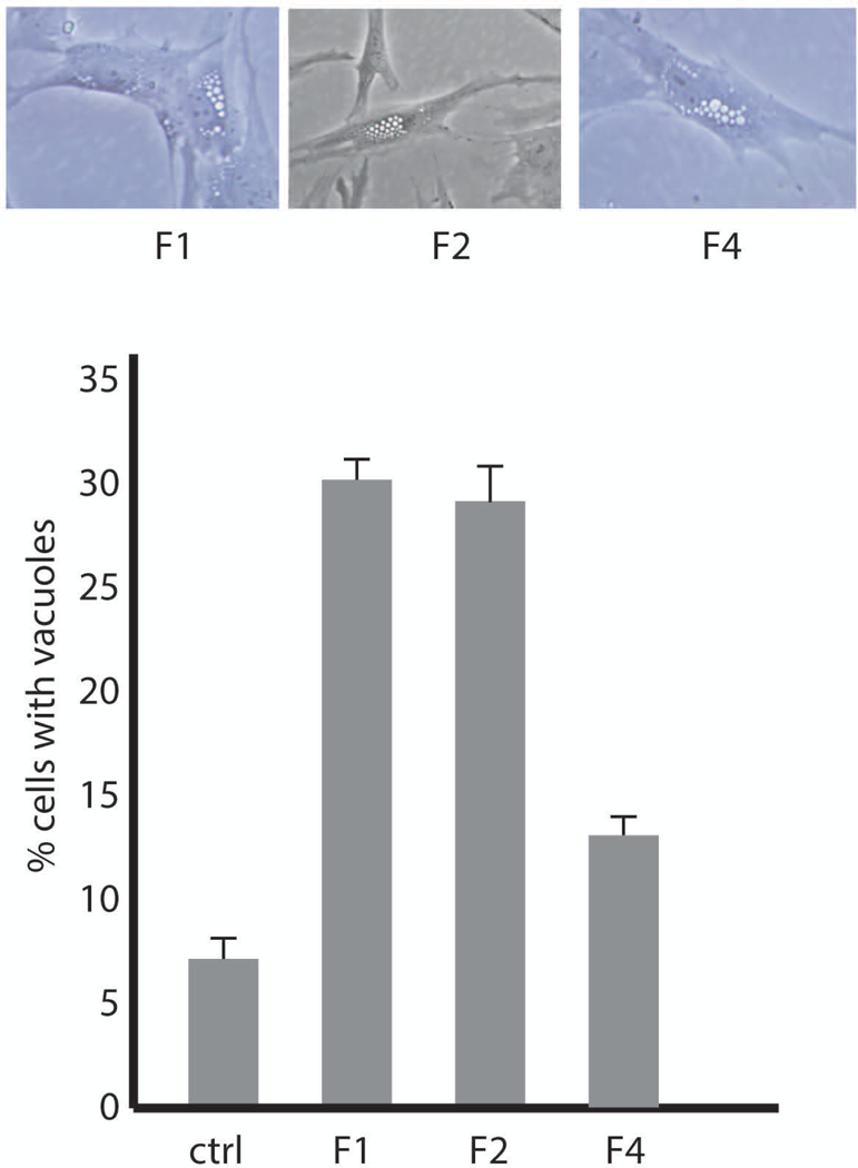
The lipid phosphatase gene FIG4 is responsible for Yunis-Varón syndrome and Charcot-Marie-Tooth disease Type 4J, a peripheral neuropathy. We now describe four families with FIG4 variants and prominent abnormalities of central nervous system (CNS) white matter (leukoencephalopathy), with onset in early childhood, ranging from severe hypomyelination to mild undermyelination, in addition to peripheral neuropathy. Affected individuals inherited biallelic FIG4 variants from heterozygous parents. Cultured fibroblasts exhibit enlarged vacuoles characteristic of FIG4 dysfunction. Two unrelated families segregate the same G > A variant in the +1 position of intron 21 in the homozygous state in one family and compound heterozygous in the other. This mutation in the splice donor site of exon 21 results in read-through from exon 20 into intron 20 and truncation of the final 115 C-terminal amino acids of FIG4, with retention of partial function. The observed CNS white matter disorder in these families is consistent with the myelination defects in the FIG4 null mouse and the known role of FIG4 in oligodendrocyte maturation. The families described here the expanded clinical spectrum of FIG4 deficiency to include leukoencephalopathy.

Keywords: CMT4J; PIKFYVE; VAC14; dysmyelination; endolysosome; leukodystrophy; neurodegeneration; oligodendrocyte; vacuolization, PtdIns(3,5)P2.


Figures

Figure 1:
Figure 1:. Evidence of CNS myelin defects in affected individuals carrying FIG4 variants. MRI images.
Axial T2 weighted MR images of patient 1(family 1) at 10 months (A,B) and 27 months (C,D) demonstrating complete absence of myelination in cerebral white matter and internal capsule with no improvement on follow up. Patient 2 (family 2) at 30 months (E,F) and 8 years (G,H) demonstrates high signal in the posterior limb of the internal capsule and diffuse high signal in the posterior periventricular and deep cerebral white matter. Family 3, sibling 1 at 25 months (I,J) demonstrates mild high signal in cerebral white matter with myelination present in deep and subcortical white matter. Family 3, sibling 2 at 18 months (K,L) demonstrates more striking hypomyelination with very little normal myelin visible. The MRI of patient 5 in family 4 at 3 years of age was largely normal with non-specific high signal in the periventricular and deep parietal white matter (M,N).
Figure 2:
Figure 2:. Recessive inheritance of FIG4 variants in four families.
(A) Affected individuals (solid symbols) are compound heterozygotes for inherited variants of FIG4. Carrier parents were unaffected (open symbols) except for the mother in Family 4 who was diagnosed with multiple sclerosis (partially filled symbol). (B) Genomic DNA sequences from affected individuals demonstrate two inherited mutant alleles. (C) Locations of variants on the FIG4 protein. Exon borders are marked. Red, VAC14 protein interaction domain; gold, phosphatase catalytic active site. FIG4 cDNA, NM_014845.5
Figure 3:
Figure 3:. Nonsense-mediated decay of the allele 1 transcript in Family 1.
The affected individual is heterozygous for variant W246X in exon 7. The sequence of the wildtype tryptophan codon TGG and the stop codon TAG are both evident in the PCR product from genomic DNA (top). In contrast, the product amplified by RT-PCR of fibroblast RNA contains the tryptophan codon TGG, but the stop codon is not detectable. The data demonstrates the instability of the stop-codon containing transcript, which is a predicted substrate for nonsense-mediated decay.
Figure 4.
Figure 4.. Retention of intron 20 in the allele 2 transcript in Family 1.
(A) location of splice site mutation at exon 21+1. (B) Aberrant, short 3’ RACE product obtained from fibroblast RNA isolated from the affected individual in Family 1 (arrow). (C) Sanger sequence of purified 3’ RACE products demonstrates correct splicing from exon 20 to exon 21 in one transcript and readthrough from exon 20 into intron 20 in the other transcript. (D) Additional sequence of the aberrant, read-through 3’ RACE product demonstrates the polyadenylation signal in intron 20, 100 bp downstream from exon 20. (E) Amino acid sequence of the truncated protein encoded by the read-through transcript.
Figure 5.
Figure 5.. Patient fibroblasts from Family 1 and Family 2 exhibit the characteristic vacuolization caused by deleterious variants of FIG4.
A) Live cell microscopy of cultured fibroblasts from affected individuals. B) Quantitation of the extent of vacuolization. Values represent means ± SD.

References

    1. Baulac S, Lenk GM, Dufresnois B, Ouled Amar Bencheikh B, Couarch P, Renard J, Larson PA, Ferguson CJ, Noe E, Poirier K, Hubans C, Ferreira S, Guerrini R, Ouazzani R, El Hachimi KH, Meisler MH, & Leguern E, (2014). Role of the phosphoinositide phosphatase FIG4 gene in familial epilepsy with polymicrogyria. Neurology, 82, 1068–1075. - PMC - PubMed
    1. Campeau PM, Lenk GM, Lu JT, Bae Y, Burrage L, Turnpenny P, Roman Corona-Rivera J, Morandi L, Mora M, Reutter H, Vulto-van Silfhout AT, Faivre L, Haan E, Gibbs RA, Meisler MH, & Lee BH, (2013). Yunis-Varon syndrome is caused by mutations in FIG4, encoding a phosphoinositide phosphatase. American journal of human genetics, 92, 781–791. - PMC - PubMed
    1. Chow CY, Zhang Y, Dowling JJ, Jin N, Adamska M, Shiga K, Szigeti K, Shy ME, Li J, Zhang X, Lupski JR, Weisman LS, & Meisler MH (2007). Mutation of FIG4 causes neurodegeneration in the pale tremor mouse and patients with CMT4J. Nature, 448, 68–72. - PMC - PubMed
    1. Corona-Rivera JR, Romo-Huerta CO, López-Marure E, Ramos FJ, Estrada-Padilla SA, Zepeda-Romero LC. (2011) New ocular findings in two sisters with Yunis-Varón syndrome and literature review. Eur J Med Genet. 54:76–81. - PubMed
    1. DiVincenzo C, Elzinga CD, Medeiros AC, Karbassi I, Jones JR, Evans MC, Braastad CD, Bishop CM, Jaremko M, Wang Z, Liaquat K, Hoffman CA, York MD, Batish SD, Lupski JR, Higgins JJ (2014). The allelic spectrum of Charcot-Marie-Tooth disease in over 17,000 individuals with neuropathy. Mol Genet Genomic Med. 2:522–529, Suppl Table 5. - PMC - PubMed

Publication types

MeSH terms