Review
. 2019 Apr;31(4):e13546.
doi: 10.1111/nmo.13546. Epub 2019 Feb 10.

Advances in the physiology of gastric emptying

Affiliations
Review

Advances in the physiology of gastric emptying

Raj K Goyal et al. Neurogastroenterol Motil. 2019 Apr.

Abstract

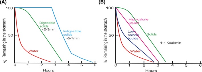
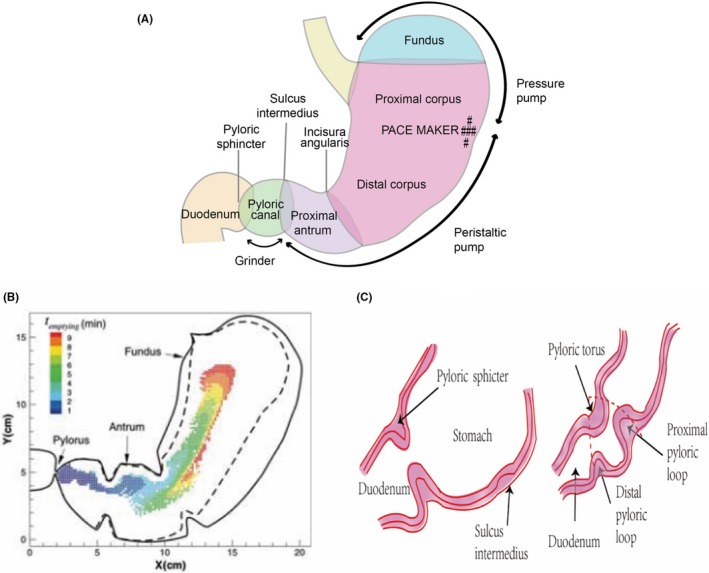
There have been many recent advances in the understanding of various aspects of the physiology of gastric motility and gastric emptying. Earlier studies had discovered the remarkable ability of the stomach to regulate the timing and rate of emptying of ingested food constituents and the underlying motor activity. Recent studies have shown that two parallel neural circuits, the gastric inhibitory vagal motor circuit (GIVMC) and the gastric excitatory vagal motor circuit (GEVMC), mediate gastric inhibition and excitation and therefore the rate of gastric emptying. The GIVMC includes preganglionic cholinergic neurons in the DMV and the postganglionic inhibitory neurons in the myenteric plexus that act by releasing nitric oxide, ATP, and peptide VIP. The GEVMC includes distinct gastric excitatory preganglionic cholinergic neurons in the DMV and postganglionic excitatory cholinergic neurons in the myenteric plexus. Smooth muscle is the final target of these circuits. The role of the intramuscular interstitial cells of Cajal in neuromuscular transmission remains debatable. The two motor circuits are differentially regulated by different sets of neurons in the NTS and vagal afferents. In the digestive period, many hormones including cholecystokinin and GLP-1 inhibit gastric emptying via the GIVMC, and in the inter-digestive period, hormones ghrelin and motilin hasten gastric emptying by stimulating the GEVMC. The GIVMC and GEVMC are also connected to anorexigenic and orexigenic neural pathways, respectively. Identification of the control circuits of gastric emptying may provide better delineation of the pathophysiology of abnormal gastric emptying and its relationship to satiety signals and food intake.

Keywords: digestive and inter-digestive periods; gastric emptying; gastric motility; intestinal hormones; neural control; satiety and food intake; the interstitial cell of Cajal; vagal circuits.


Conflict of interest statement

The authors report no conflict of interest relevant to this article.

Figures

Figure 1
Figure 1
Gastric emptying rates vary with the physical characteristic and caloric density of food. (A) Effect of physical characteristics of food on the rate of gastric emptying. Note that water or 5% glucose leaves the stomach at a fast rate, and digestible solids begin to leave after a lag period and leave the stomach slowly. Large pieces of indigestible solids are retained in the stomach during the digestive period and are then rapidly emptied. (B) Effect of caloric density of the liquid meal. Note that water leaves the stomach very fast and only 50% remains in the stomach at 10 min. High‐calorie liquids empty at a slower rate with 50% remaining in the stomach at 2 h. Low‐calorie liquids empty at an intermediate rate so the 50% leave the stomach by 1 h
Figure 2
Figure 2
Anatomic and functional parts of the human stomach, the gastric tunnel (Magenstrasse), and the pylorus. (A) Anatomic and functional parts of the stomach. The stomach includes three multifunctional, interconnected structures: pressure pump, peristaltic pump, and a grinder. The pressure pump includes anatomic fundus and proximal corpus. The peristaltic pump includes anatomic distal corpus and pyloric antrum. The pressure and peristaltic pumps form the propulsive unit. The anatomic correlate of the grinder is the pylorus that includes the anatomic pyloric canal and pyloric sphincter. Modified from Adler.15 (B) A functional tunnel along the lesser curvature of the stomach, called Magenstrasse, that may allow liquids to bypass the slower movement of the solid food through the stomach to accomplish a very fast gastric emptying. The figure identifies the initial location of particles emptied during 10 min, gray shaded with the time period of emptying, t emptying. From Pal et al10 (C) Details of the pyloric complex which includes the proximal muscle loop and the distal muscle loop formed by the pyloric sphincter. The proximal and distal muscle loops are ~2 cm away from each other on the greater curvature but merge together on the lesser curvature of the stomach. The loops enclose a triangular cavity with the merged muscle loops forming a torus at the lesser curve. The pyloric torus fits into the groove left between the proximal and distal muscle loops along the greater curvature, like a pastel and mortar, to form a perfect grinder. Pylorus provides mechanical grinding and food that has been tenderized by acid‐pepsin, to form chyme. The proximal muscle loop and the pyloric sphincter are separately regulated and can work independently
Figure 3
Figure 3
A simplified gastric inhibitory vagal circuit (GIVC) and the gastric excitatory vagal circuit (GEVC). (A) The GIVC includes GIVMC and its inputs. GIVMC consists of preganglionic DMV‐C‐i neuron and postganglionic, NANC inhibitory neuron in the myenteric plexus (MP‐NANC‐i). See text for details of the neurotransmission. The DMV‐C‐i neurons receive excitatory input directly from the NTS‐CC‐i neurons via the α1‐receptors and through NTS‐PPG neurons via GHSR or GLP‐1 receptors. The NTS‐CC‐i neurons receive glutaminergic input from low‐threshold vagal afferents whose neurons are in the nodose ganglion (NG). (Arrow—stimulation; flat—inhibition). (B) The GEVC includes GEVMC and its inputs. GEVMC consists of preganglionic DMV‐C‐e neurons and postganglionic, cholinergic excitatory myenteric plexus (MP‐C‐e) neurons. DMV‐C‐e neurons receive strong inhibitory input from NTS‐GABA‐e neurons and NTS‐CC‐e neurons, and excitatory input from NTS‐GLUT‐e neurons. The NTS‐GABA‐e, NTS‐CC‐e, and NTS‐GLUT‐e neurons are interconnected and send integrated inhibitory input to the DMV‐C‐e neurons. NTS‐CC neurons also send inhibitory input to DMV‐C‐e neurons via the α2‐receptors. The inhibitory inputs from the NTS to DMV‐C‐e suppress spontaneously active DMV‐C‐e and cause gastric relaxation. On the other hand, suppression of the NTS‐GABA neurons NTS‐CC‐i disinhibits the spontaneously active DMV‐C‐e neurons leading to gastric excitation and fast gastric emptying as in acute hypoglycemia. See text for other details. (Arrow—stimulation; flat—inhibition)
Figure 4
Figure 4
Main sites of action of CCK and ghrelin. (A) Cholecystokinin (CCK) is a prototype breaking hormone. It acts to stimulate vagovagal circuit at multiple levels. CCK stimulates vagal afferents endings by paracrine effect and enhances glutamate release from the vagal afferent endings projecting onto NTS‐CC‐i neurons. CCK also directly or indirectly stimulates NTS‐CC‐i neurons, PPG‐i neurons, and NTS‐POMC‐S neurons. CCK stimulation of NTS‐CC‐i neurons activates DMV‐C‐i via the α1‐adrenergic receptor; stimulation of the NTS‐PPG‐i neurons via the GLP‐1 receptor on the DMV‐C‐i. CCK has also been shown to directly stimulate MP‐NANC‐i neurons, and may also stimulate DMV‐C‐i neurons. Thus, CCK acts at multiple sites to stimulate GIVC. Stimulation of NTS‐CC‐i neurons also inhibits DMV‐C‐e neurons via the α2‐adrenergic receptors. Thus, CCK also acts to inhibit GEVC. All these actions further augment the inhibitory effect of CCK on the gastric muscle. CCK also stimulates NTS‐POMC‐s neurons to generate satiety signals. (Arrow—stimulation; flat—inhibition). (B) Ghrelin is a gastric accelerating hormone. Ghrelin acts at multiple central and peripheral sites to stimulate gastric motility. Centrally, ghrelin inhibits NTS‐CC‐i neurons thereby inhibiting DMV‐C‐i. Ghrelin also inhibits NTS‐CC‐e to disinhibit DMV‐C‐e neurons. Ghrelin also disinhibits DMV‐C‐e neurons by inhibiting AP‐GABA‐e neurons that project onto DMV‐C‐e neurons. Ghrelin also acts on myenteric plexus and the smooth muscle. All these actions lead to strong gastric excitation. (Arrow—stimulation; flat—inhibition)

References

    1. Kar P, Jones KL, Horowitz M, Chapman MJ, Deane AM. Measurement of gastric emptying in the critically ill. Clin Nutr. 2015;34(4):557‐564. - PubMed
    1. Ehrlein HJ, Schemann J. Gastrointestinal Motility. Munich: Technische Universität München; 2005.
    1. Horowitz M, Dent J. Disordered gastric emptying: mechanical basis, assessment and treatment. Baillieres Clin Gastroenterol. 1991;5(2):371‐407. - PubMed
    1. Hunt JN, Smith JL, Jiang CL. Effect of meal volume and energy density on the gastric emptying of carbohydrates. Gastroenterology. 1985;89(6):1326‐1330. - PubMed
    1. Meyer JH, Elashoff J, Porter‐Fink V, Dressman J, Amidon GL. Human postprandial gastric emptying of 1‐3‐millimeter spheres. Gastroenterology. 1988;94(6):1315‐1325. - PubMed

Publication types