Review
. 2019 Jul 15;20(14):1739-1753.
doi: 10.1002/cbic.201900017. Epub 2019 May 24.

Small-Molecule Inhibitors of the Proteasome's Regulatory Particle

Affiliations
Review

Small-Molecule Inhibitors of the Proteasome's Regulatory Particle

Christine S Muli et al. Chembiochem. .

Abstract

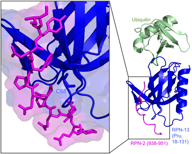
Cells need to synthesize and degrade proteins consistently. Maintaining a balanced level of protein in the cell requires a carefully controlled system and significant energy. Degradation of unwanted or damaged proteins into smaller peptide units can be accomplished by the proteasome. The proteasome is composed of two main subunits. The first is the core particle (20S CP), and within this core particle are three types of threonine proteases. The second is the regulatory complex (19S RP), which has a myriad of activities including recognizing proteins marked for degradation and shuttling the protein into the 20S CP to be degraded. Small-molecule inhibitors of the 20S CP have been developed and are exceptional treatments for multiple myeloma (MM). 20S CP inhibitors disrupt the protein balance, leading to cellular stress and eventually to cell death. Unfortunately, the 20S CP inhibitors currently available have dose-limiting off-target effects and resistance can be acquired rapidly. Herein, we discuss small molecules that have been discovered to interact with the 19S RP subunit or with a protein closely associated with 19S RP activity. These molecules still elicit their toxicity by preventing the proteasome from degrading proteins, but do so through different mechanisms of action.

Keywords: 19S RP; cancer; inhibitors; proteasome; ubiquitin.


Figures

Figure 1.
Figure 1.
Cryo-electron microscopy (cryo-EM) structure of the 26S proteasome that was released in 2016. (PDB code: 5GJR)
Figure 2.
Figure 2.
The ubiquitination cascade starts with the E1 enzyme, which activates ubiquitin (Ub). E1 then passes activated ubiquitin to the E2 enzyme. The E2-ubiquitin complex can then interacts with an E3, which is the ubiquitin ligase. This complex can transfer ubiquitin to the substrate (S). This process is repeated until the substrate has a polyubiquitin chain of 4-5 ubiquitin moieties. This chain can then be recognized by the 19S RP of the proteasome to begin degradation of the substrate.
Figure 3.
Figure 3.
Structures of 20S CP inhibitors. (A) Bortezomib is a tripeptide that reversibly inhibits β5 catalytic activity utilizing a boronic acid moiety. (B) Carfilzomib is a tetrapeptide that irreversibly inhibits β5 catalytic activity via an epoxyketone.
Figure 4.
Figure 4.
(A). Schematic of Rpn-13’s interactions with Uch37, Ub, and Rpn-2. (B) Solution structure of full-length Rpn-13 solved in 2010, with the DEUBAD (orange) and Pru (blue) domain indicated. (PDB: 2KR0)
Figure 5.
Figure 5.
Close-up of the Pru domain Rpn-13:Rpn-2 interface, with Cys-88 (RA-190’s binding residue) highlighted. (PDB: 5V1Y)
Figure 6.
Figure 6.
(A) Cryo-EM structure showing Usp14’s and UbAl’s position when docked onto the 19S RP. (PDB: 5GJQ) (B) Co-crystal of USP14’s catalytic domain and IU1 demonstrating the binding pocket of IU1. (PDB: 6IIK).
Figure 7.
Figure 7.
X-ray crystal structure close-up of S. cerevisiae Rpn-11:Rpn-8 heterodimer to display the Zn2+ ion site on Rpn-11. (PDB: 4O8X)
Figure 8.
Figure 8.
(A) Cryo-EM structure of heterohexameric ATPase ring of the Rpt subunits (PDB: 5GJQ) (B) Cryo-EM structure of the homohexameric ATPase ring of p97, with one protomer highlighted (above) and the D1, D2, and N domains indicated (below). (PDB: 5FTK)
Figure 9.
Figure 9.
There remains a large number of 19S RP subunits that that may be amenable to inhibition by small molecules. Additionally, preventing PTMs or designing PROTACs to overexpressed subunits may also be viable methods to disrupt proteasome activity.

References

    1. Dikic I, Annu. Rev. Biochem. 2017, 86, 193. - PubMed
    1. Lee DH, Goldberg AL, Trends Cell Biol. 1998, 8, 397. - PubMed
    1. Lecker SH, J. Am. Soc. Nephrol. 2006, 17, 1807. - PubMed
    1. Etlinger JD, Goldberg AL, Proc. Natl. Acad. Sci. 1977, 74, 54. - PMC - PubMed
    1. Goldberg AL, Dice JF, Annu. Rev. Biochem. 1974, 43, 835. - PubMed

Publication types

MeSH terms

Substances

LinkOut - more resources