. 2019 Feb 19;8(4):e009960.
doi: 10.1161/JAHA.118.009960.

MG 53 Protein Protects Aortic Valve Interstitial Cells From Membrane Injury and Fibrocalcific Remodeling

Affiliations

MG 53 Protein Protects Aortic Valve Interstitial Cells From Membrane Injury and Fibrocalcific Remodeling

T M Ayodele Adesanya et al. J Am Heart Assoc. .

Erratum in

Abstract

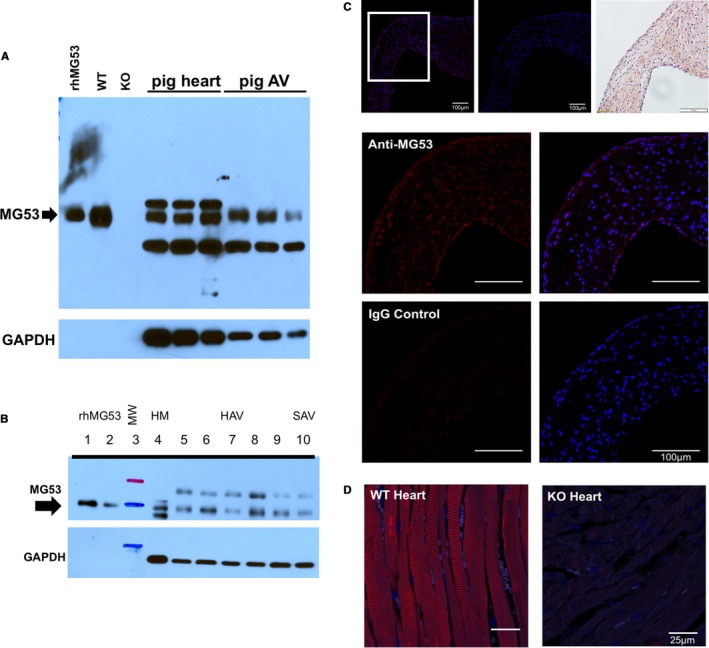
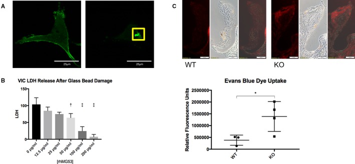
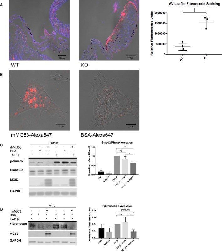
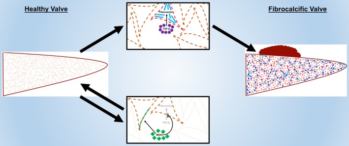
Background The aortic valve of the heart experiences constant mechanical stress under physiological conditions. Maladaptive valve injury responses contribute to the development of valvular heart disease. Here, we test the hypothesis that MG 53 (mitsugumin 53), an essential cell membrane repair protein, can protect valvular cells from injury and fibrocalcific remodeling processes associated with valvular heart disease. Methods and Results We found that MG 53 is expressed in pig and human patient aortic valves and observed aortic valve disease in aged Mg53-/- mice. Aortic valves of Mg53-/- mice showed compromised cell membrane integrity. In vitro studies demonstrated that recombinant human MG 53 protein protects primary valve interstitial cells from mechanical injury and that, in addition to mediating membrane repair, recombinant human MG 53 can enter valve interstitial cells and suppress transforming growth factor-β-dependent activation of fibrocalcific signaling. Conclusions Together, our data characterize valve interstitial cell membrane repair as a novel mechanism of protection against valvular remodeling and assess potential in vivo roles of MG 53 in preventing valvular heart disease.

Keywords: cell membrane repair; fibrosis; heart valve; transforming growth factor‐β; valvular heart disease.


Figures

Figure 1
Figure 1
MG53 is expressed in pig and human patient aortic valve tissue. A, Western blotting shows that MG53 (53 kDa, arrow) is expressed in pig myocardium and aortic valves. rhMG53 (0.03 ng) and wild‐type mouse myocardium (0.06 μg) were used as positive controls and Mg53−/− myocardium (0.06 μg) as a negative control. 10 μg of lysates from pig tissues were loaded from 3 different animals. The apparent lack of GAPDH expression in the mouse myocardial tissue reflects the ≈150‐fold less loading of mouse vs pig protein lysates. B, Western blotting shows that MG53 (53 kDa, arrow) is expressed in human myocardium and aortic valves. rhMG53 was loaded in lanes 1 to 2 (0.05 and 0.02 ng, respectively); ladder in lane 3; non‐failing myocardium (2.5 μg) in lane 4; non‐diseased valves (10 μg) in lanes 5 to 9; and a stenotic valve (10 μg) in lane 10. C, Immunohistochemistry of axial sections of pig aortic valve shows that MG53 (red) is expressed in the valve interstitial layers as well as its endothelial linings. In the top‐most row, ×20 magnification images are shown of (left to right) MG53 staining, rabbit immunoglobulin G control staining, and hematoxylin and eosin staining. In the lower rows and from the ×20 boxed area, ×40 magnification images are shown of MG53 and rabbit immunoglobulin G control staining (red) and overlaid DAPI (4′,6‐diamidino‐2‐phenylindole) staining (blue). D, MG53 staining (red) of wild‐type and Mg53−/− mouse myocardium are positive and negative controls, respectively. AV indicates aortic valve; HAV, healthy aortic valves; HM, healthy myocardium; IgG, immunoglobulin G; KO, knockout (Mg53−/−); MW, molecular weight; SAV, stenotic aortic valve; rh, recombinant human; WT, wild‐type.
Figure 2
Figure 2
Welcome3jah3Aged Mg53−/− mice develop aortic valve disease. A, Peak aortic valve outflow velocities were measured via echocardiography with higher velocities associated with presence of aortic stenosis. Ten‐ and 24‐month‐old wild‐type mice had no statistical difference in peak aortic valve velocities. Twenty‐four‐month‐old Mg53−/− mice showed statistically significant higher velocities compared with both 10‐month‐old Mg53−/− mice and 24‐month‐old wild‐type mice. A 2‐way ANOVA test with Tukey multiple comparison testing (α=0.05) was used to obtain P values. n=9 ten‐month‐old wild‐type mice; n=6 ten‐month‐old Mg53−/− mice; n=14 twenty‐four‐month‐old wild‐type mice; n=6 twenty‐four‐month‐old Mg53−/− mice. B, Representative color Doppler images and pulse‐wave aortic valve jets are shown for wild‐type (left) and Mg53−/− (right) mice. Aortic valve disease is associated with more turbulent color images and higher velocities as measured by the y‐axis of the pulse‐wave jets. C, Coronal sections of aortic valve leaflets are shown opening upwards, left ventricle below and aorta above. Histologic analyses of aged wild‐type (left) and Mg53−/− (right) mouse hearts using hematoxylin and eosin staining showed increased area; (D) Masson trichrome staining showed increased fibrosis; and (E) Alizarin Red S staining showed increased calcification in Mg53−/− aortic valve leaflets (n=4) compared with those from wild‐type control mice (n=4). Mann–Whitney test was used to obtain the P value for (C), and 2‐tailed Student t tests were used to obtain P values for (D and E). AV indicates aortic valve; KO, knockout (Mg53−/−); WT, wild‐ type. *P<0.05, **P<0.01, ****P<0.0001.
Figure 3
Figure 3
MG53 protects against valve interstitial cell (VIC) membrane injury. A, In pig VICs transfected with GFPMG53 (left), microelectrode needle penetration of the VIC membrane causes translocation of GFPMG53 to the membrane injury site (right, boxed; n=10 cells). B, VICs were exposed to physical damage via vigorous shaking with glass micro‐beads. rhMG53 reduces LDH release—an index of cell membrane injury—in a dose‐dependent manner (n=3 biological replicates). An ordinary, 1‐way ANOVA test with Dunnett multiple comparison testing (α=0.05) was used to obtain P values. The ANOVA P‐value was P<0.0001. Multiple comparison testing compared each rhMG53‐treatment group to the control, non‐treated group to obtain P values <0.05 for cells treated with 50, 100, and 200 μg/mL rhMG53. C, EBD is impermeable to cells with intact membranes. Aortic valve leaflets of 24‐month‐old Mg53−/− mice (n=4) show significantly increased EBD uptake compared with those of littermate wild‐type mice (n=3). Representative images are shown on top, and relative fluorescence quantification is shown below. A 2‐tailed Student t test was used to obtain the P‐value. These results suggest increased endothelial permeability in 24‐month‐old Mg53−/− compared with wild‐type controls. Further, given that EBD will not penetrate cells with intact plasma membranes, these results also suggest a greater in vivo burden of cell membrane injury in aortic valves from Mg53−/− mice. Imaging shows increased damage particularly in endothelial and subendothelial cell layers. KO indicates knockout (Mg53−/−); LDH, lactate dehydrogenase; rh, recombinant human; WT, wild‐type. *P<0.05, **P<0.01, ***P<0.001.
Figure 4
Figure 4
rhMG53 suppresses TGF‐β‐induced fibrocalcific signaling in valve interstitial cells. A, Immunohistochemistry of aged wild‐type (left) and Mg53−/− (right) mouse hearts show increased fibronectin staining in Mg53−/− aortic valve leaflets (n=3) compared with those from wild‐type control mice (n=4). A 2‐tailed Student t test was used to obtain the P value. In different tissues, fibronectin mediates both fibrosis and calcification such as that observed in histologic analysis of 24‐month‐old Mg53−/− mouse valves (Figure 2).41, 42, 43, 44 Fibronectin is a downstream product of the TGF‐ß pathway and has been associated with VIC injury, facilitating continued signaling events and VIC activation.7, 8, 45, 48, 49, 50, 57 Given the relationship between TGF‐ß and fibronectin and with increased fibronectin staining in Mg53−/− aortic valves, we aimed to target our in vitro studies through our histopathology findings. Specifically, we tested if rhMG53 treatment could modulate VIC TGF‐ß signaling and downstream fibronectin expression. B, To guide our mechanistic insights, we first wanted to determine if rhMG53 could enter pig aortic VICs. Here, we conjugated rhMG53 and BSA as a control to the Alexa‐647 fluorophore to allow visualization of rhMG53‐specific cell entry via live cell imaging (n=10 cells). Importantly, rhMG53 VIC entry was observed after treating cells for 30 minutes, thus our use of this pretreatment time in our in vitro signaling studies. C, Short time points to study cell signaling events in pig VICs showed that rhMG53 reduces TGF‐β‐induced Smad2 phosphorylation (n=3). D, Longer time points to study differences in protein expression in pig VICs demonstrated that rhMG53 reduces TGF‐β‐induced fibronectin expression (n=4). Ordinary, 1‐way ANOVA tests with Tukey multiple comparison testing (α=0.05) were used to obtain P‐values. AV indicates aortic valve; BSA, bovine serum albumin; KO, knockout (Mg53−/−); rh, recombinant human; TGF, transforming growth factor; WT, wild type. *P<0.05, **P<0.01, ***P=0.001.
Figure 5
Figure 5
A suggested model summarizes our results. Accumulated membrane injury to VICs (dashed lines) is associated with increased TGF‐β‐induced fibronectin expression, allowing for pooled TGF‐β signaling, constitutive VIC activation (red lines), and downstream fibrocalcific changes. These remodeling processes result in VHD. MG53 protects VICs from both membrane injury (green patches) and fibrocalcific TGF‐β signaling to prevent such remodeling and maintain valvular homeostasis. Future studies will specify mechanisms of MG53‐mediated protection and will assess the translational potential for MG53‐based protein therapies or MG53‐coated cardiovascular materials in preventing or treating VHD.

References

    1. Thanassoulis G. Lipoprotein (a) in calcific aortic valve disease: from genomics to novel drug target for aortic stenosis. J Lipid Res. 2016;57:917–924. - PMC - PubMed
    1. Rajamannan NM, Gersh B, Bonow RO. Calcific aortic stenosis: from bench to the bedside—emerging clinical and cellular concepts. Heart. 2003;89:801–805. - PMC - PubMed
    1. Lester WM, Gotlieb AI. In vitro repair of the wounded porcine mitral valve. Circ Res. 1988;62:833–845. - PubMed
    1. Lester WM, Damji AA, Tanaka M, Gedeon I. Bovine mitral valve organ culture: role of interstitial cells in repair of valvular injury. J Mol Cell Cardiol. 1992;24:43–53. - PubMed
    1. Lester WM, Damji AA, Gedeon I, Tanaka M. Interstitial cells from the atrial and ventricular sides of the bovine mitral valve respond differently to denuding endocardial injury. In Vitro Cell Dev Biol. 1993;29A:41–50. - PubMed

Publication types

MeSH terms

LinkOut - more resources