. 2019;9(1):97-107.
doi: 10.3233/JPD-181443.

HSP90 and Its Novel Co-Chaperones, SGT1 and CHP-1, in Brain of Patients with Parkinson's Disease and Dementia with Lewy Bodies

Affiliations

HSP90 and Its Novel Co-Chaperones, SGT1 and CHP-1, in Brain of Patients with Parkinson's Disease and Dementia with Lewy Bodies

Anastasiia Bohush et al. J Parkinsons Dis. 2019.

Abstract

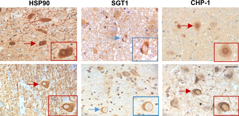
Background: Parkinson's disease (PD) is a neurodegenerative disorder characterized by the presence of inclusions known as Lewy bodies in some brain regions. Lewy bodies consist of α-synuclein and many other proteins including chaperones.

Objective: To learn more about the role of chaperone complexes in PD and a related disorder, i.e., dementia with Lewy bodies (DLB), in this work we analyzed the expression of HSP90 and its two quite recently identified co-chaperones, SGT1 and CHP-1, in selected brain regions from patients suffering from these diseases.

Methods: To fulfill the aim of our study we used human material and applied immunohistochemistry, Western blot analysis and real time/quantitative PCR (RT-qPCR).

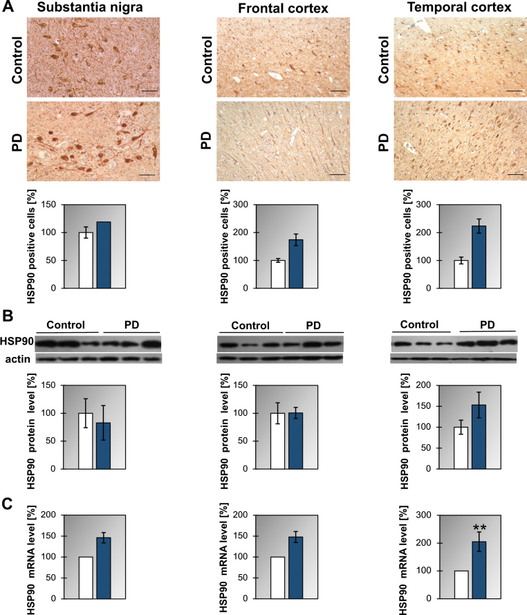
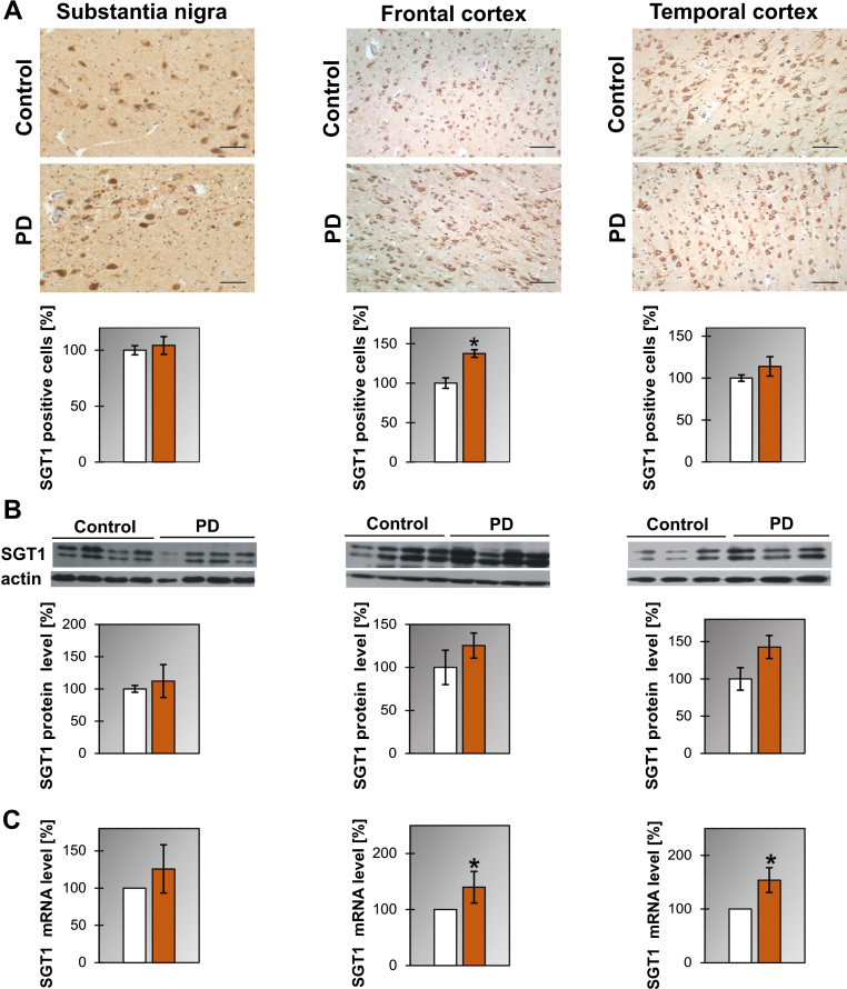
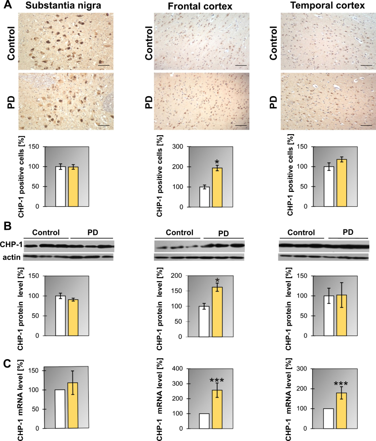
Results: We have found that HSP90 mRNA level is higher in the temporal cortex of PD and in frontal cortex of DLB brains, even though level of protein does not change significantly. The mRNA level of SGT1 is higher in the frontal and temporal cortex of PD and in substantia nigra of DLB brains while no significant changes in the level of protein were noticed. Similarly, the mRNA level of CHP-1 was found to be higher in the frontal and temporal cortex of PD and in all examined regions i.e. substantia nigra, frontal and temporal cortex of DLB brains. In the case of CHP-1 the protein level was found to be higher in frontal cortex of PD and in all examined areas of DLB patients.

Conclusions: Our data indicate that the level of HSP90, SGT1 and CHP-1 is upregulated in the majority of cases of PD and DLB, which suggests that the examined proteins might be involved in these pathologies.

Keywords: CHP-1; HSP90; Parkinson’s disease; SGT1; dementia with Lewy bodies.


Conflict of interest statement

The authors have no conflict of interest to report.

Figures

Fig.1
Fig.1
Presence of HSP90 in the substantia nigra, frontal and temporal cortex. (A) Representative immunohistochemical images from the control and PD group are shown. Scale bar is 60μm. Lower panel presents the analysis of HSP90 positive neurons. (B) Western blot (upper panel) and densitometric analysis of HSP90 level (lower panel). (C) RT-qPCR of HSP90 mRNA level. In (B) and (C) samples from 3 control and 3 PD patients were analyzed. White and blue bars represent control and PD cases, respectively. Error bars indicate mean±SEM; **p < 0.01.
Fig.2
Fig.2
Presence of SGT1 in the substantia nigra, frontal and temporal cortex. (A) Representative immunohistochemical images from the control and PD group are shown. Scale bar is 60μm. Lower panel presents the analysis of SGT1 positive neurons. (B) Western blot (upper panel) and densitometric analysis of SGT1 level (lower panel). (C) RT-qPCR of SGT1 mRNA level. In (B) and (C) samples from 3-4 control and 3-4 PD patients were analyzed. White and orange bars represent control and PD cases, respectively. Error bars indicate mean±SEM; *p < 0.05.
Fig.3
Fig.3
Presence of CHP-1 in the substantia nigra, frontal and temporal cortex. (A) Representative immunohistochemical images from the control and PD group are shown. Scale bar is 60μm. Lower panel presents the analysis of CHP-1 positive neurons. (B) Western blot (upper panel) and densitometric analysis of CHP-1 level (lower panel). (C) RT-qPCR of CHP-1 mRNA level. In (B) and (C) samples from 3 control and 3 PD patients were analyzed. White and yellow bars represent control and PD cases, respectively. Error bars indicate mean±SEM; *p < 0.05; ***p < 0.001.
Fig.4
Fig.4
Localization of HSP90, SGT1 and CHP-1 in Lewy bodies in the substantia nigra of PD (upper panel) and DLB brain (lower panel). Scale bar is 50μm.

References

    1. Lan D, Wang W, Zhuang J, Zhao Z (2015) Proteasome inhibitor-induced autophagy in PC12 cells overexpressing A53T mutant α-synuclein. Mol Med Rep 11, 1655–1660. - PMC - PubMed
    1. Ziemssen T, Reichmann H (2007) Non-motor dysfunction in Parkinson’s disease. Parkinsonism Relat Disord 13, 323–332. - PubMed
    1. Bohnen NI, Albin RL (2011) The cholinergic system and Parkinson disease. Behav Brain Res 221, 564–573. - PMC - PubMed
    1. Przedborski S (2017) The two-century journey of Parkinson disease research. Nat Rev Neurosci 18, 251–259. - PubMed
    1. Surmeier DJ, Obeso JA, Halliday GM (2017) Selective neuronal vulnerability in Parkinson disease. Nat Rev Neurosci 18, 101–113. - PMC - PubMed

Publication types