Review
. 2019 Feb 11;10(2):123.
doi: 10.1038/s41419-019-1385-8.

Neurotrophins and their involvement in digestive cancers

Affiliations
Review

Neurotrophins and their involvement in digestive cancers

Sabrina Blondy et al. Cell Death Dis. .

Abstract

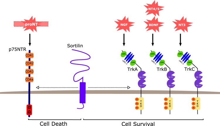
Cancers of the digestive system, including esophageal, gastric, pancreatic, hepatic, and colorectal cancers, have a high incidence and mortality worldwide. Efficient therapies have improved patient care; however, many challenges remain including late diagnosis, disease recurrence, and resistance to therapies. Mechanisms responsible for these aforementioned challenges are numerous. This review focuses on neurotrophins, including NGF, BDNF, and NT3, and their specific tyrosine kinase receptors called tropomyosin receptor kinase (Trk A, B, C, respectively), associated with sortilin and the p75 neurotrophin receptor (p75NTR), and their implication in digestive cancers. Globally, p75NTR is a frequently downregulated tumor suppressor. On the contrary, Trk and their ligands are considered oncogenic factors. New therapies which target NT and/or their receptors, or use them as diagnosis biomarkers could help us to combat digestive cancers.


Conflict of interest statement

The authors declare that they have no conflict of interest.

Figures

Fig. 1
Fig. 1. The fate of NT receptors’ family and cells following ligand binding.
Trk Tropomyosin Receptor Kinase, NGF Neuronal Growth Factor, BDNF Brain-derived Neurotrophic Factor, NT Neurotrophin, CRD Cystein-Rich Domain, LRR Leucine-Rich motif, IgL Immunoglobulin-Like domain, TKD Tyrosin Kinase Domain, DD Death Domain
Fig. 2
Fig. 2. mNT/Trk pro-survival signaling involving the RAS-MAPK, PI3K/AKT, PLC gamma (γ) downstream pathways.
mNT homodimers bind to their respective Trk and induce dimerization and transphosphorylation at tyrosine residues of the TKD located in the cytoplasmic tails. This phosphorylation allows the binding of sh2 domain-mediated adaptor proteins including (i) the p85-PI3K subunit, (ii) PLCγ and (iii) Grb2/SOS. (i) PI3K mediates AKT phosphorylation and activation, through PDK1. P-AKT phosphorylates IκB, triggering its ubiquitinylation and subsequent proteasome degradation, leading to NF-κB transcription factor release. (ii) PLCγ converts PI(4,5)P2 into IP3 and DAG, allowing the activation of PKC activation and, subsequently, MAPK pathways (RAF/RAF-MEK-ERK 1, 2). P-ERK 1,2 can activate IkB, which activated NF-κB, and its translocation into nucleus to activate transcription factors and/or display transcription factor functions. Simultaneously, IP3 can also trigger intracellular Ca2+ release, leading to the activation of NF-κB as well as NFAT, after its dephosphorylation through calmodulin/calcineurin. (iii) Finally, Grb2/SOS also activates MAPK pathways, similarly to PLCγ-PKC. All these pathways trigger transcription of genes involved in cell survival, proliferation, migration, invasion, angiogenesis, and EMT, leading to tumor growth and maintenance. Trk Tropomyosin Receptor Kinase, NT Neurotrophin, mNT mature Neurotrophin, CDR Cystein-Rich Domain, LRR Leucine-Rich motif, IgL Immunoglobulin-Like domain, TKD Tyrosin Kinase Domain, PI3K phosphatidylinositol-3-kinase, PKC Protein Kinase C, PLC Phospholipase C, PDK Phosphoinositide-Dependent Kinase, P phosphorylated residues, Ub Ubiquitin residues, DAG Diacylglycerol, IP3 Inositol Triphosphate, PI(4,5)P2 Phosphatidylinositol 4,5-biphosphate, MAPK Mitogen-Activated Protein Kinase, NFAT Nuclear Factor of Activated T-cells, EMT Epithelial-Mesenchymal Transition
Fig. 3
Fig. 3. Balance between pro-apoptotic and pro-survival signaling induced by p75NTR, modulated by sortilin and the nature of binding of NT (mNT or proNT).
a ProNT binding to the complex sortilin/ p75NTR triggers the recruitment of adaptator proteins, including TRAF6, NRAGE, and NRIF, to the p75NTR intracellular DD. These proteins activate MEKK, leading to the phosphorylation of both p38 and JNK pathways, with p62 and c-Jun, p53 activation, respectively. Altogether, these three proteins promote a decreased expression of the anti-apoptotic protein Bcl-2, concomitant with an increased expression of pro-apoptotic proteins such as Bax, Bad, and Bim. Bax translocates into the mitochondria membrane, thus forming a complex with BAK, which triggers both CytC and AIF release. Together with P-c-Jun, AIF translocate into the nucleus and induces the transcription of pro-apoptotic genes, as well as DNA fragmentation, both leading to cell death. At the same time, CytC forms the apoptosome complex with the procaspase 9 and Apaf-1, allowing caspase 9 activation (cleavage) and subsequent caspase 3 activation. Finally, the caspase 3 cleaves its substrate, the PARP, which translocates into the nucleus to exert similar effects on cell death as described above. b p75NTR can be cleaved by α-secretases, hence releasing its extracellular domain (p75NTR-ECD) which can bind both mNT and proNT in the extracellular media. The resting cleaved fragment anchored to cell membrane (p75NTR-CTF) and complexed to sortilin, is sufficient to induce death signaling through the same mechanisms as described above. p75NTR can also be cleaved by γ-secretase in its intracellular part, leading to the release of its intracellular domain (p75NTR-ICD) containing the DD. p75NTR-ICD can translocate into the nucleus to induce cell death but mechanisms are currently poorly understood. c On the contrary, after mNT binding, the p75-ICD fragment is released, as well as the CTF or full-length form of this receptor bound to the intracellular adaptator protein TRAF6 and complexed to sortilin, and can induce cell survival through the same NF-κB signaling as described previously. Trk Tropomyosin Receptor Kinase, NT Neurotrophin, mNT mature Neurotrophin, p75NTR p75 neurotrophin receptor, CDR Cystein-Rich Domain, LRR Leucine-Rich motif, IgL Immunoglobulin-Like domain, TKD Tyrosin Kinase Domain, DD Death Domain, P phosphorylated residues, Ub Ubiquitin residues, ECD Extracellular Domain, CTF C-Terminal Fragment, ICD Intracellular Domain, TRAF6 TNF Receptor associated Factor 6, NRIF Neurotrophin Receptor-Interacting Factor, NRAGE Neurotrophin Receptor-interacting MAGE homolog, MEKK MAP Kinase Kinase or MAP3K, JNK c-Jun N-terminal Kinase, Bax Bcl-2-associated X, BAD Bcl-2-associated Death promoter, Bak Bcl-2-homologous antagonist/killer, cytC: cytochrome C, PARP Poly(ADP-Ribose) Polymerase

Similar articles

Cited by

References

    1. Bothwell, M. Recent advances in understanding neurotrophin signaling. F1000Research5, 1885 (2016). - PMC - PubMed
    1. Glerup S, Nykjaer A, Vaegter CB. Sortilins in neurotrophic factor signaling. Handb. Exp. Pharmacol. 2014;220:165–189. doi: 10.1007/978-3-642-45106-5_7. - DOI - PubMed
    1. Huang EJ, Reichardt LF. Trk receptors: roles in neuronal signal transduction. Annu. Rev. Biochem. 2003;72:609–642. doi: 10.1146/annurev.biochem.72.121801.161629. - DOI - PubMed
    1. Reichardt LF. Neurotrophin-regulated signalling pathways. Philos. Trans. R. Soc. B Biol. Sci. 2006;361:1545–1564. doi: 10.1098/rstb.2006.1894. - DOI - PMC - PubMed
    1. Meldolesi J. Neurotrophin receptors in the pathogenesis, diagnosis and therapy of neurodegenerative diseases. Pharmacol. Res. 2017;121:129–137. doi: 10.1016/j.phrs.2017.04.024. - DOI - PubMed

Publication types

MeSH terms