

Flavones hydroxylated at 5, 7, 3' and 4' ameliorate skin fibrosis via inhibiting activin receptor-like kinase 5 kinase activity
- PMID: 30741930
- PMCID: PMC6370799
- DOI: 10.1038/s41419-019-1333-7
Flavones hydroxylated at 5, 7, 3' and 4' ameliorate skin fibrosis via inhibiting activin receptor-like kinase 5 kinase activity
Abstract
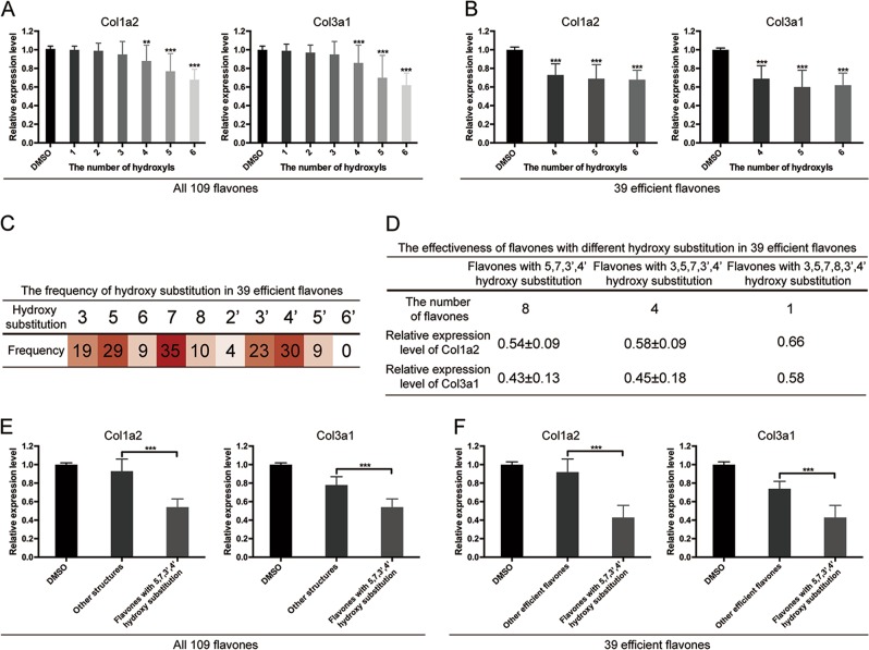
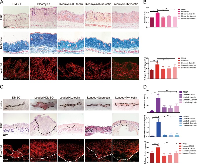
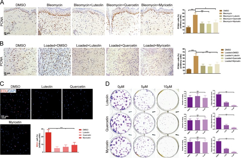
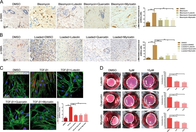
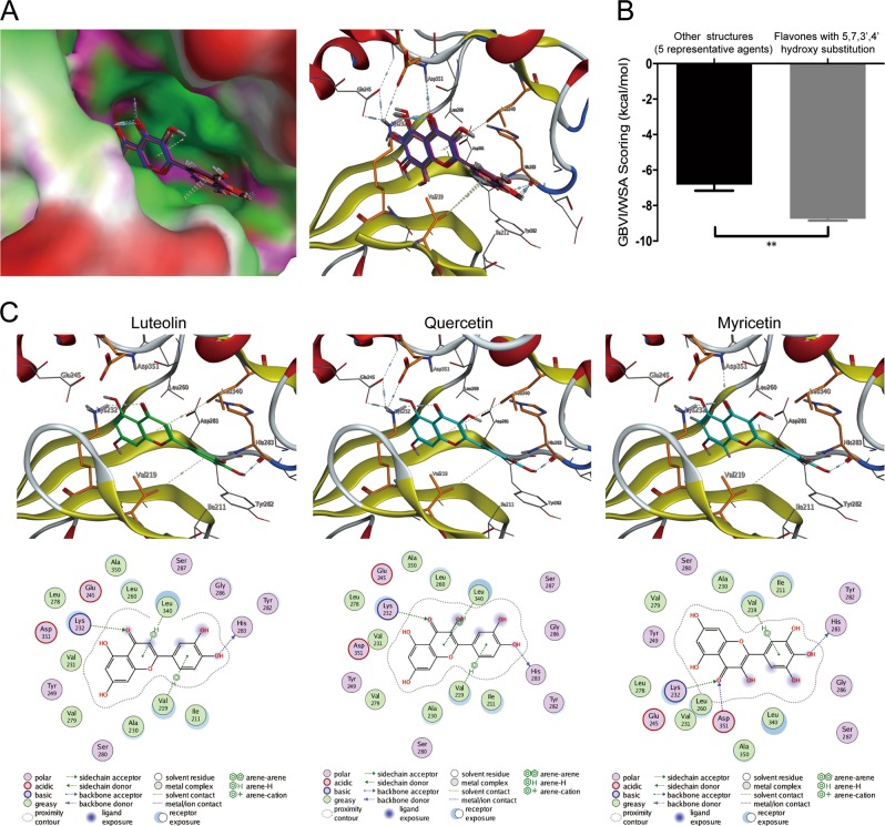
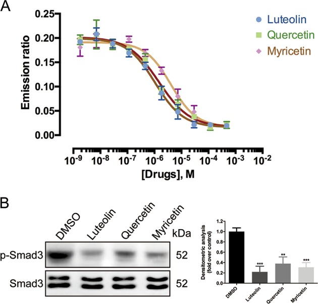
Skin fibrosis is mainly characterized by excessive collagen deposition. Studies have recently identified a number of flavonoids with variable structures that have the potency of inhibiting collagen synthesis and thus attenuating organ fibrosis. In this study, we found that flavones with 5, 7, 3', 4' hydroxy substitution reduced collagen expression most efficiently. Among those flavones, luteolin, quercetin, and myricetin were selected for follow-up. In vivo, the three compounds ameliorated skin fibrosis and reduced collagen deposition. Further analysis showed the compounds had significant inhibition on the proliferation, activation and contractile ability of dermal fibroblasts in vitro and in vivo. More importantly, we revealed that luteolin, quercetin, and myricetin selectively downregulated the phosphorylation of Smad2/3 in TGF-β/Smads signaling via binding to activin receptor-like kinase 5 (ALK5) and impairing its catalytic activity. We also found flavones with 5, 7, 3', 4' hydroxy substitution showed stronger affinity with ALK5 compared with other flavonoids. Herein, we identified at least in part the underlying molecular basis as well as the critical structures that contribute to the antifibrotic bioactivity of flavones, which might benefit drug design and modification.
Conflict of interest statement
The authors declare that they have no conflict of interest.
Figures

Similar articles
-
Galangin inhibits hypertrophic scar formation via ALK5/Smad2/3 signaling pathway.Mol Cell Biochem. 2016 Feb;413(1-2):109-18. doi: 10.1007/s11010-015-2644-3. Epub 2016 Jan 4. Mol Cell Biochem. 2016. PMID: 26728998
-
Dihydromyricetin attenuates hypertrophic scar formation by targeting activin receptor-like kinase 5.Eur J Pharmacol. 2019 Jun 5;852:58-67. doi: 10.1016/j.ejphar.2019.02.039. Epub 2019 Feb 23. Eur J Pharmacol. 2019. PMID: 30807748
-
Inhibition of activin receptor-like kinase 5 attenuates bleomycin-induced pulmonary fibrosis.Exp Mol Pathol. 2007 Aug;83(1):39-46. doi: 10.1016/j.yexmp.2006.12.003. Epub 2006 Dec 24. Exp Mol Pathol. 2007. PMID: 17274978
-
Intracellular TGF-beta receptor blockade abrogates Smad-dependent fibroblast activation in vitro and in vivo.J Invest Dermatol. 2006 Aug;126(8):1733-44. doi: 10.1038/sj.jid.5700303. Epub 2006 Jun 1. J Invest Dermatol. 2006. PMID: 16741519
-
Baicalein attenuates hypertrophic scar formation via inhibition of the transforming growth factor-β/Smad2/3 signalling pathway.Br J Dermatol. 2016 Jan;174(1):120-30. doi: 10.1111/bjd.14108. Epub 2015 Nov 8. Br J Dermatol. 2016. PMID: 26301336
Cited by
-
The role of heat shock protein 90 in idiopathic pulmonary fibrosis: state of the art.Eur Respir Rev. 2025 Mar 19;34(175):240147. doi: 10.1183/16000617.0147-2024. Print 2025 Jan. Eur Respir Rev. 2025. PMID: 40107664 Free PMC article. Review.
-
The potential of functionalized dressing releasing flavonoids facilitates scar-free healing.Front Med (Lausanne). 2022 Oct 3;9:978120. doi: 10.3389/fmed.2022.978120. eCollection 2022. Front Med (Lausanne). 2022. PMID: 36262272 Free PMC article. Review.
-
Laricitrin 3-Rutinoside from Ginkgo biloba Fruits Prevents Damage in TNF-α-Stimulated Normal Human Dermal Fibroblasts.Antioxidants (Basel). 2023 Jul 15;12(7):1432. doi: 10.3390/antiox12071432. Antioxidants (Basel). 2023. PMID: 37507970 Free PMC article.
-
Curcumin and its nano-formulations: Defining triple-negative breast cancer targets through network pharmacology, molecular docking, and experimental verification.Front Pharmacol. 2022 Aug 8;13:920514. doi: 10.3389/fphar.2022.920514. eCollection 2022. Front Pharmacol. 2022. PMID: 36003508 Free PMC article.
-
Inhibitory Effects of 3',4'-Dihydroxyflavonol in a Mouse Model of Glaucoma Filtration Surgery and TGFβ1-Induced Responses in Human Tenon's Fibroblasts.Transl Vis Sci Technol. 2022 Aug 1;11(8):18. doi: 10.1167/tvst.11.8.18. Transl Vis Sci Technol. 2022. PMID: 35980669 Free PMC article.
References
Publication types
MeSH terms
Substances
LinkOut - more resources
Full Text Sources
Research Materials
