. 2019 Feb 11;9(1):1742.
doi: 10.1038/s41598-018-38264-1.

Molecular layer interneurons shape the spike activity of cerebellar Purkinje cells

Affiliations

Molecular layer interneurons shape the spike activity of cerebellar Purkinje cells

Amanda M Brown et al. Sci Rep. .

Abstract

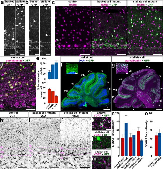
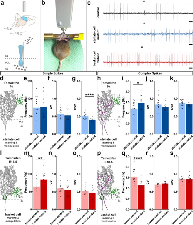
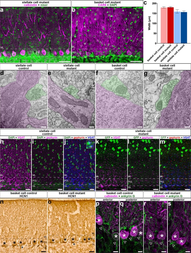
Purkinje cells receive synaptic input from several classes of interneurons. Here, we address the roles of inhibitory molecular layer interneurons in establishing Purkinje cell function in vivo. Using conditional genetics approaches in mice, we compare how the lack of stellate cell versus basket cell GABAergic neurotransmission sculpts the firing properties of Purkinje cells. We take advantage of an inducible Ascl1CreER allele to spatially and temporally target the deletion of the vesicular GABA transporter, Vgat, in developing neurons. Selective depletion of basket cell GABAergic neurotransmission increases the frequency of Purkinje cell simple spike firing and decreases the frequency of complex spike firing in adult behaving mice. In contrast, lack of stellate cell communication increases the regularity of Purkinje cell simple spike firing while increasing the frequency of complex spike firing. Our data uncover complementary roles for molecular layer interneurons in shaping the rate and pattern of Purkinje cell activity in vivo.


Conflict of interest statement

The authors declare no competing interests.

Figures

Figure 1
Figure 1
The Ascl1CreER allele can be used for genetic marking of stellate cells and basket cells. (a) Schematic of cerebellar circuitry. The Purkinje cell (PC), basket cell (BC), and stellate cell (SC) are colorized while other cells and fibers in the cerebellar cortex are represented in grayscale (climbing fiber = CF, mossy fiber = MF, Golgi cell = GoC, granule cell = GrC, unipolar brush cell = UBC). Dotted lines represent the borders of the Purkinje cell layer (PCL) with the molecular layer (ML) and granule cell layer (GL). (b,c) Golgi-Cox stain of cerebellar tissue. Scale = 50 μm. (b) Basket cell (arrowhead) and Purkinje cell (asterisk) revealed by Golgi-Cox stain. (c) Stellate cell (arrowhead) and Purkinje cells (asterisks) revealed by Golgi-Cox stain. (d) Representation of breeding scheme. (e) Schematic of methods for tamoxifen administration. Tamoxifen was administered via oral gavage to pregnant dams at E18.5 to achieve constitutive marking and manipulation of a subset of basket cells in the resulting pups (upper left). Tamoxifen was administered via subcutaneous injection into the scruff of pups at P4 to achieve constitutive marking and manipulation of a subset of stellate cells (bottom right). (f) Labeled cells were found in the basal molecular layer in animals treated with tamoxifen at the basket cell timepoint and the apical molecular layer for those treated at the stellate cell timepoint (g). Scale = 50 μm. 5 sections separated by ~200 µm around midline per mouse, N = 7 for each condition.
Figure 2
Figure 2
Ascl1CreER conditional deletion of VGAT protein is efficient and selective. (a,b) Labeled cells in the basket and stellate conditions. Scale = 20 μm. 5 sections separated by ~200 µm around midline were analyzed per mouse, N = 7 for each condition. (a) Labeled basket cells (left) and labeled stellate cells (right) in customary regions of the ML. (b) Labeled basket cells (left) and stellate cells (right) farther from their traditional regions of the ML. (c) RORα expression in the ML of control (left), basket cell mutant (middle), and stellate cell mutant (right) mice. Scale = 50 μm. Per condition: N = 3, n = 9. (d) Representation of recombination quantification. GFP-expressing cells were counted (yellow circles) and compared to a count of the total number of ML interneurons. Scale = 50 μm. (e) Quantification of recombination efficiency in basket and stellate cell conditions. (f,g) Sample of a whole sagittal cerebellar section in the basket (f) and stellate (g) cell manipulation conditions. White boxes indicate the regions that are shown as blow-ups in the insets. Cerebellar lobules are indicated with Roman numerals. Scale = 0.5 mm. Inset scale = 50 μm. (h–j) VGAT expression across the ML in control mice (h), basket cell VGAT deletion mice (i) and stellate cell VGAT deletion mice (j). Scale = 50 μm. (k–m) Putative synapses where VGAT and GFP overlapped (arrowheads) could be readily found in control tissue (k), but were absent or not readily found in the apical molecular layer of mutant stellate cell tissue (l) as well as the basal molecular layer of mutant basket cell tissue (m). Scale = 10 μm. Control N = 3, n ≥ 3; basket cell mutant: N = 2, n ≥ 3; stellate cell mutant: N = 6, n ≥ 3. (n) Quantification of VGAT puncta density in the basal, middle, and apical ML of basket and stellate VGAT mutant mice. (o) Quantification of VGAT puncta density in the entire ML in both mutant conditions. (a–d,h–j) Dotted lines indicate the borders of the Purkinje cell layer (PCL) with the molecular layer (ML) above and the granular layer (GL) below.
Figure 3
Figure 3
Genetic depletion of GABAergic molecular layer interneuron neurotransmission alters Purkinje cell firing in vivo. (a) Schematic of electrophysiology setup. ML (molecular layer), PCL (Purkinje cell layer), GL (granular layer) (b) Picture of a mouse in the electrophysiology setup. (c) Example recordings of Purkinje cells in a control (top), stellate cell mutant (middle), and basket cell mutant (bottom) mouse. Complex spikes indicated with asterisks. Scale = 20 ms. (d) Schematic of a stellate cell (green) in relation to a Purkinje cell (grey). (e–g) Purkinje simple spike electrophysiology in the stellate cell condition (control: N = 7, n = 20; mutant: N = 3, n = 15). (e,f) Firing frequency (e) and CV (f) were not significantly different. (g) CV2 was significantly decreased compared to control. (h) Schematic of a climbing fiber (magenta) to a stellate cell (green) and a Purkinje cell (grey). (i–k) Purkinje complex spike electrophysiology in the stellate cell condition (control: N = 7, n = 20; mutant: N = 3, n = 15). (i) Firing frequency was significantly increased compared to control. (j,k) Neither CV (j) nor CV2 (k) were significantly different. (l) Schematic of a basket cell (green) in relation to a Purkinje cell (grey). (m–o) Purkinje simple spike electrophysiology in the basket cell condition (control: N = 5, n = 17; mutant: N = 3, n = 18). (m) Firing frequency was significantly increased. (n,o) Neither CV (n) nor CV2 (o) were significantly changed. (p) Schematic of a climbing fiber (magenta) in relation to a basket cell (green) and a Purkinje cell (grey). (q–s) Purkinje cell complex spike electrophysiology in the basket cell condition (control: N = 5, n = 17; mutant: N = 3, n = 18). (q) Firing frequency was significantly decreased in mutants compared to control mice. (r,s) Neither CV (r) nor CV2 (s) were significantly changed.
Figure 4
Figure 4
Deleting Vgat in molecular layer interneurons does not rearrange cerebellar circuitry or induce neurodegeneration. (a,b) Examples of images used for quantification of ML thickness. Purkinje cell layer (PCL), molecular layer (ML), and granular layer (GL). Scale = 50 μm. (c) Quantification of ML thickness in all conditions (basket cell control mean = 179.9 μm ± 3.83, N = 3, n = 9, basket cell mutant mean = 181.1 μm ± 3.16, N = 3, n = 9, P = 0.82; stellate cell control mean = 159.7 μm ± 9.20, N = 3, n = 18, stellate cell mutant mean = 157.2 μm ± 5.50, N = 6, n = 36, P = 0.83). (d,e) TEM images revealed normal synapses in all conditions. Purkinje cells and processes are colorized in magenta and putative basket and stellate synaptic terminals are colorized in green. Scale = 200 nm. (d,e) Stellate cell: N = 2, n ≥ 10, per condition. (f,g) Basket cell: N = 3, n ≥ 9, per condition. (h–m) Gephyrin expression was unchanged in stellate cell mutant mice compared to control. Scale = 20 μm. (h–j) Control mice (N = 3, n ≥ 3) have uniform expression of VGAT (h,j) and gephyrin (i,j) in the ML. (j) Example triple labeled synapses (arrowhead) are shown in the blowup. (k–m) Stellate cell mutant mice do not have uniform expression of VGAT (k). However, gephyrin appears uniformly expressed (l). (m) We did not detect triple stained synapses. N = 3, n ≥ 9. (n–s) HCN1 staining suggests the region of the basket cell pinceau is unchanged from control (n) in basket cell mutant mice (o). Scale = 20 μm. N = 4, n ≥ 12. (p–s) The Purkinje cell axon initial segment (arrowheads) is obvious in control (p,q) and basket cell mutant mice (r–s) throughout the cerebellum with example images shown from both anterior and posterior lobules. Purkinje cell somas are indicated by asterisks. Scale = 10 μm. Per condition N = 3, n ≥ 9. (a,b,h–s) Dotted lines indicate the borders of the Purkinje cell layer (PCL) with the molecular layer (ML) above and the granular layer (GL) below.
Figure 5
Figure 5
Conditional deletion of Vgat in molecular layer interneurons does not lead to gross cerebellar changes in cellular composition, cellular distribution, or layer patterning. (a–x) Cerebellar cell types were present and appeared unchanged in location and morphology despite the lack of VGAT in basket cells and stellate cells. Dotted lines indicate the borders of the Purkinje cell layer (PCL) with the molecular layer (ML) and the granular layer (GL). Scale = 20 μm. Control: N = 14, n ≥ 42; basket cell condition: N = 4, n ≥ 12; stellate cell condition: N = 6, n ≥ 18. (a–c) CAR8 and IP3R1. (d–f) GABAαR6. (g–i) NFH. (j–l) Neurogranin. (m–o) VGLUT1. (p–r) VGLUT2. (s–u) Calretinin. (v–x) CART.
Figure 6
Figure 6
Conditional deletion of Vgat with Ascl1CreER occurs in extracerebellar cell types that are unlikely to affect Purkinje cell activity in this manipulation. (a) Sparse labeling of cells occurs outside the cerebellum at the basket cell time point. Scale = 1 mm. (b–g) Many of the cells outside the cerebellum had morphologies that resembled glia, with the notable exception of cells in the olfactory bulb (g) where the majority of cells had the morphology of neurons, though cells with glial-like morphology were also present. Scale = 20 μm. (h) Sparse labeling outside of the cerebellum also occurred at the stellate cell time point. Scale = 1 mm. (i–n) While again many of the cells had morphologies that resembled glia, some cells with neuron-like morphologies were also detected. (k) Very sparse labeling of putative granule cells in the dentate gyrus occurred at the stellate cell time point, unlike at the basket cell time point at which no neurons were detected in the hippocampus. (n) Similar to the basket cell time point, many neurons in the olfactory bulb were labeled in addition to some glial-like cells. Scale = 20 μm. (o) Recombined cells with glial-like morphologies co-labeled with GFP and CAII, a maker of oligodendrocytes. Scale = 20 μm. N = 2, n ≥ 6. (p) Cells with neuron-like morphologies in the dentate gyrus of the hippocampus co-labeled with GFP and NeuN. Scale = 20 μm. N = 2, n ≥ 6. (q) Both neurons (arrow, co-labeled with GFP and NeuN) and glia (arrowhead, only labeled with GFP and not by NeuN) were labeled in the olfactory bulb. Scale = 20 μm. N = 2, n ≥ 6.

Similar articles

Cited by

References

    1. Manto M, et al. Consensus Paper: Roles of the Cerebellum in Motor Control—The Diversity of Ideas on Cerebellar Involvement in Movement. The Cerebellum. 2012;11:457–487. doi: 10.1007/s12311-011-0331-9. - DOI - PMC - PubMed
    1. Eccles JC. Circuits in the cerebellar control of movement. Proc. Natl. Acad. Sci. USA. 1967;58:336–43. doi: 10.1073/pnas.58.1.336. - DOI - PMC - PubMed
    1. Reeber, S. L., Otis, T. S. & Sillitoe, R. V. New roles for the cerebellum in health and disease. Front Syst Neurosci. 2013 Nov 14,7,83, 10.3389/fnsys.2013.00083, eCollection (2013). - PMC - PubMed
    1. Voogd J, Glickstein M. The anatomy of the cerebellum. Trends Neurosci. 1998;21:370–5. doi: 10.1016/S0166-2236(98)01318-6. - DOI - PubMed
    1. Eccles JC, Llinás R, Sasaki K. Parallel fibre stimulation and the responses induced thereby in the Purkinje cells of the cerebellum. Exp. brain Res. 1966;1:17–39. - PubMed

Publication types

MeSH terms

Substances

LinkOut - more resources