Comparative Study
. 2019 Jun;13(6):1469-1483.
doi: 10.1038/s41396-019-0361-8. Epub 2019 Feb 11.

Comparative cytology, physiology and transcriptomics of Burkholderia insecticola in symbiosis with the bean bug Riptortus pedestris and in culture

Affiliations
Comparative Study

Comparative cytology, physiology and transcriptomics of Burkholderia insecticola in symbiosis with the bean bug Riptortus pedestris and in culture

Tsubasa Ohbayashi et al. ISME J. 2019 Jun.

Abstract

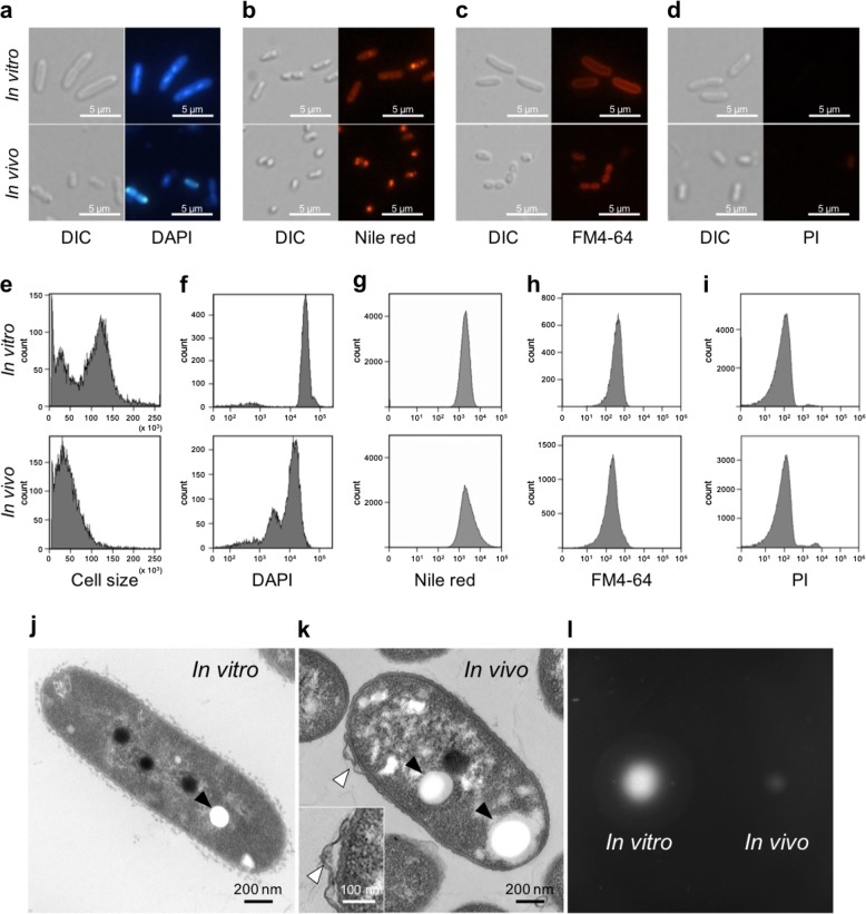
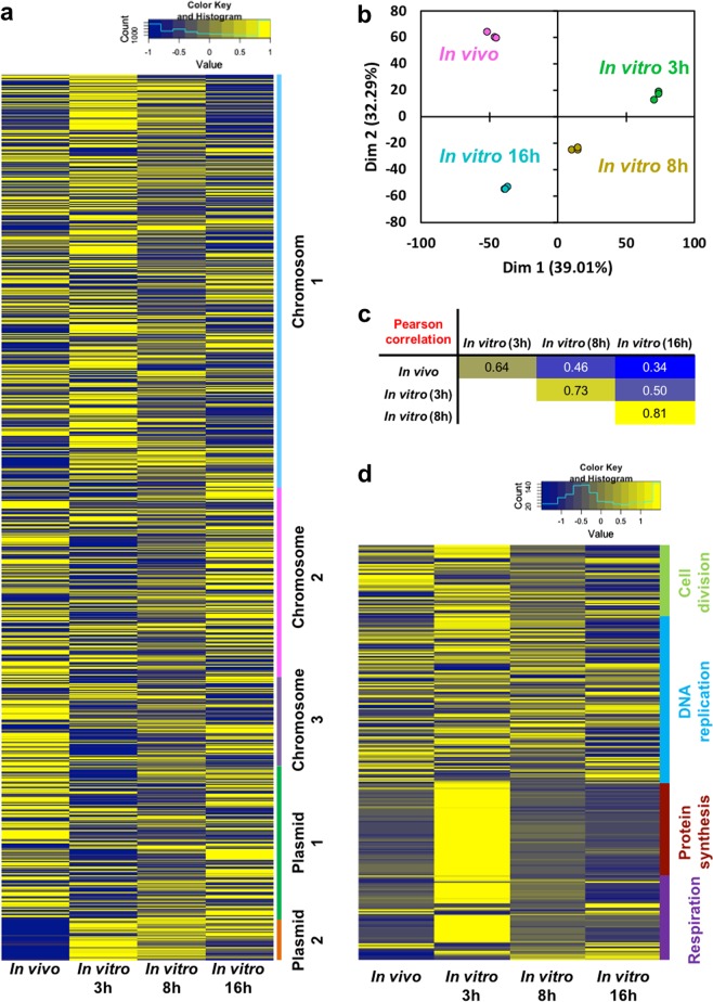
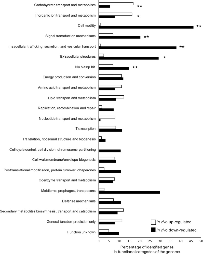
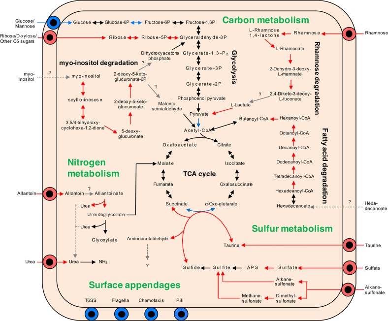
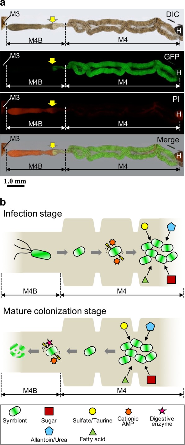
In the symbiosis of the bean bug Riptortus pedestris with Burkholderia insecticola, the bacteria occupy an exclusive niche in the insect midgut and favor insect development and reproduction. In order to understand how the symbiotic bacteria stably colonize the midgut crypts and which services they provide to the host, we compared the cytology, physiology, and transcriptomics of free-living and midgut-colonizing B. insecticola. The analyses revealed that midgut-colonizing bacteria were smaller in size and had lower DNA content, they had increased stress sensitivity, lost motility, and an altered cell surface. Transcriptomics revealed what kinds of nutrients are provided by the bean bug to the Burkholderia symbiont. Transporters and metabolic pathways of diverse sugars such as rhamnose and ribose, and sulfur compounds like sulfate and taurine were upregulated in the midgut-colonizing symbionts. Moreover, pathways enabling the assimilation of insect nitrogen wastes, i.e. allantoin and urea, were also upregulated. The data further suggested that the midgut-colonizing symbionts produced all essential amino acids and B vitamins, some of which are scarce in the soybean food of the host insect. Together, these findings suggest that the Burkholderia symbiont is fed with specific nutrients and also recycles host metabolic wastes in the insect gut, and in return, the bacterial symbiont provides the host with essential nutrients limited in the insect food, contributing to the rapid growth and enhanced reproduction of the bean bug host.


Conflict of interest statement

The authors declare that they have no conflict of interest.

Figures

Fig. 1
Fig. 1
Bacterial cell morphology of in vivo and in vitro Burkholderia symbiont cells. ad Differential interference contrast (left) and fluorescence microscopy (right) images of in vitro and in vivo bacteria stained with DAPI a, Nile red b, FM4-64 c, and PI d. ei Flow cytometry analysis of in vivo and in vitro symbiont cells measuring cell size by light scatter e, DNA content by DAPI staining f, PHA accumulation by Nile red staining g, and membrane area by FM4-64 staining h, membrane permeability by PI staining i. j, k Transmission electron microscopy images of an in vitro j and an in vivo k symbiont cell. Filled arrows indicate PHA granules and open arrows shows a membrane bleb. l Motility test of in vitro (left) and in vivo (right) cells (see also Supplementary Fig. S3)
Fig. 2
Fig. 2
Comparison of stress sensitivities between in vivo and in vitro bacteria. The in vivo and in vitro Burkholderia symbiont cells were exposed to a SDS, b tween 20, c protease K, d LL-37, e polymyxin B, f riptocin, g CCR0008, h CCR0179, i CCR0480, j hydrogen peroxide, k sodium chloride, l glucose, and m ethanol at the indicated concentrations. The relative growth (%) was normalized by setting untreated bacterial growth to 100%. Mean ± SD (n = 3) is shown in in vitro (solid bar) and in vivo cells (open bar). Statistically significant differences between in vivo and in vitro symbiont cells were analyzed by Student’s t-test with Bonferroni correction: *P < 0.05. CFU data of the stress tests are shown in Supplementary Table S3
Fig. 3
Fig. 3
Gene expression profiles of in vivo and in vitro Burkholderia symbiont. a heatmap of 5872 gene expression profiles, b Principal component analysis, and c the pairwise Pearson correlation coefficients of in vivo and 3 h-, 8 h-, and 16 h-cultured in vitro cells. In the heatmap, the normalized mean expression level of the three biological replicates was used, and the color scale from blue to yellow indicates the relative expression level. d Gene expression profile of core cellular functions. The heatmap shows the expressions of 281 genes encoding cell division, DNA replication, protein synthesis, and respiration in in vivo and in vitro symbiont cells
Fig. 4
Fig. 4
COG functional classification of in vivo upregulated and down-regulated genes. The in vivo upregulated and down-regulated genes were classified using the NCBI COG 2014 database. Asterisks indicate statistically significant difference between in vivo and in vitro cells (Fisher’s exact test with Bonferroni correction; *P < 0.05, **P < 0.01)
Fig. 5
Fig. 5
Overview of KEGG metabolic pathways in in vivo upregulated and down-regulated genes. Arrows indicate metabolic direction, and the color shows gene expression levels in RNA-seq: red, in vivo upregulatd; blue, in vivo down-regulated; black, un-changed expression levels
Fig. 6
Fig. 6
A hypothetical model for the life cycle of the Burkholderia symbiont in the midgut of R. pedestris. a The PI staining in the midgut of 5th instar nymph infected with the strain RPE225 (a GFP-expressing mutant). DIC, GFP fluorescence, PI fluorescence, and merged images are shown. Arrows indicate the border of GFP and PI fluorescent signals. M3, midgut 3rd section; M4, midgut 4th section (crypts); M4B, M4 bulb; H, hindgut. A red auto-fluorescence was observed in M3 and H. b Graphical summary of Burkholderia features and midgut functions of R. pedestris. (Upper) During the infection stage of Burkholderia symbiont at the insect midgut, the Burkholderia symbiont cells lose flagellar motility and modify their envelope under the influence of host stress factors such as cationic AMPs (e.g. CCRs), they proliferate by metabolizing host waste materials, such as sulfate and allantoin. (Lower) In the mature colonization stage, these symbiont cells are digested in the M4B section mediated by host factors such as cathepsin proteases and CCRs, and the host absorbs nutrients derived from whole-bacterial cells

References

    1. Schwartzman JA, Ruby EG. Stress as a normal cue in the symbiotic environment. Trends Microbiol. 2016;24:414–24. - PMC - PubMed
    1. Yang DC, Blair KM, Salama NR. Staying in shape: the impact of cell shape on bacterial survival in diverse environments. Microbiol Mol Biol Rev. 2016;80:187–203. - PMC - PubMed
    1. Buchner P. Endosymbiosis of animals with plant microorganims. New York: Interscience; 1965.
    1. Bourtzis K, Miller T. Insect symbiosis. Boca Raton, FL: CRC Press; 2003.
    1. Kikuchi Y. Endosymbiotic bacteria in insects: their diversity and culturability. Microbes Environ. 2009;24:195–204. - PubMed

Publication types

MeSH terms