. 2019 Mar;20(3):362-372.
doi: 10.1038/s41590-018-0305-x. Epub 2019 Feb 11.

Mosaic nanoparticle display of diverse influenza virus hemagglutinins elicits broad B cell responses

Affiliations

Mosaic nanoparticle display of diverse influenza virus hemagglutinins elicits broad B cell responses

Masaru Kanekiyo et al. Nat Immunol. 2019 Mar.

Erratum in

Abstract

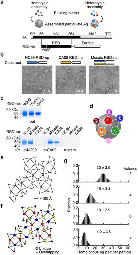
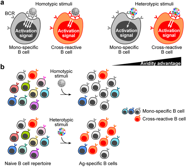
The present vaccine against influenza virus has the inevitable risk of antigenic discordance between the vaccine and the circulating strains, which diminishes vaccine efficacy. This necessitates new approaches that provide broader protection against influenza. Here we designed a vaccine using the hypervariable receptor-binding domain (RBD) of viral hemagglutinin displayed on a nanoparticle (np) able to elicit antibody responses that neutralize H1N1 influenza viruses spanning over 90 years. Co-display of RBDs from multiple strains across time, so that the adjacent RBDs are heterotypic, provides an avidity advantage to cross-reactive B cells. Immunization with the mosaic RBD-np elicited broader antibody responses than those induced by an admixture of nanoparticles encompassing the same set of RBDs as separate homotypic arrays. Furthermore, we identified a broadly neutralizing monoclonal antibody in a mouse immunized with mosaic RBD-np. The mosaic antigen array signifies a unique approach that subverts monotypic immunodominance and allows otherwise subdominant cross-reactive B cell responses to emerge.


Figures

Fig. 1.
Fig. 1.. Design and characterization of a mosaic array of heterotypic antigens on self-assembling nps.
(a) Design of HA RBD-np. Alteration in the residue 98 (Y98F) was made to abrogate sialic acid-binding property of HA. SP, signal peptide; T/C, transmembrane/cytoplasmic domains. (b) Negative-stain EM images of self-assembled HA RBD-nps. RBD-np were made using either single building blocks (left and middle) or two different building blocks (right). Shown are representative images from one experiment. (c) Antigenic characterization of RBD-np by immunoprecipitation (IP). The mAbs 3u-u (anti-NC99), 2D1 (anti-CA09) and C179 (anti-HA stem) were used to pull-down NC99, CA09 RBDs, and HA stem (control), respectively. Similar results were obtained from two independent experiments. (d) Schematic model of ferritin-based np. Twenty-four spatially dispersed antigens (colored individually) are displayed on the surface. Positions 2–6 are localized within a 100 Å distance from position 1. (e) Ferritin snub cube net with positions numbered similarly to panel d. Connected lines indicate adjacent positions located within 50–100 Å. (f) Twenty-four positions are colored according with the chromatic number (three) to avoid the same color from being located within 100 Å radius of each other. (g) Simulated likelihood of homologous antigen pairs made within 100 Å radius on a single particle by using 2, 4, 6 or 8 different building blocks (valence).
Fig. 2.
Fig. 2.. Immune induction by HA RBD-nps displaying homogeneous- and mosaic antigen arrays.
(a) Antibody responses to homologous NC99 virus. Serum HAI and pseudotype neutralization (PN) IC50 titers are measured after 2 immunizations of indicated valences of either admixed or mosaic RBD-nps with Sigma adjuvant system. For HAI, cumulative data of 3 independent experiments (n = 15) except for 8-valent admix group (2 independent experiments, n = 10) are shown. For PN, shown are representative data from one experiment (n = 5). Experiments are independently performed 3 times (2 times for 8-valent admix group) with similar results. Lines indicate means. Statistical analyses are done with one-way ANOVA with Tukey’s multiple comparisons post-hoc test. Non-statistical significance between groups is not shown for clarity. (b) Microneutralization (MN) IC50 titers against 5 different viruses in mice after priming with TIV and after boosting with mosaic RBD-np. Immune sera were collected at week 5 (prime) and 12 (boost). MN IC50 titers against each virus is plotted on five axes and connected by a line (n = 10). MN IC50 across 5 H1N1 viruses is shown as GMT ± geometric s.d. Statistical analyses are done with two-tailed paired t-test. Data are from one experiment. (c) Schematic representation of different immunization modalities. (d) Serum HAI titers to NC99 virus after 2 (NC99, admix and mosaic groups) or 4 (sequential) immunizations with adjuvant. Shown are representative data from one experiment (n = 10). Experiments are independently performed two times with similar results. Statistical analysis is done with one-way ANOVA with Tukey’s multiple comparison post-hoc test. (e) MN IC50 titers against 5 H1N1 viruses. MN IC50 titers are plotted as b. Shown are representative data from one experiment (n = 10). Experiments are independently performed two times with similar results. Statistical analysis is done with one-way ANOVA with Tukey’s multiple comparisons post-hoc test.
Fig. 3.
Fig. 3.. Induction of cross-reactive HA-specific B cells by RBD-nps.
(a) Gating strategy for identifying HA-specific B cells by flow cytometry. Anti-CD3, CD8, CD14, and F4/80 were combined and used to separate T cells, monocytes, and macrophages (MØ). A disparate pair of HAs (NC99 and CA09) was used to define cross-reactivity of HA-specific B cells. Frequencies of IgD B cells specific to NC99 HA (b), CA09 (c), or cross-reactive to NC99 and CA09 (d) in PBMCs of mice immunized with different RBD-nps (n = 15, cumulative data of 3 independent experiments except for 8-valent admix group (n = 10, cumulative of 2 independent experiments). Statistical analyses are done with one-way ANOVA with Tukey’s multiple comparisons post-hoc test by using valence = 1 as a comparator. (e) Mutation rate in IGHV genes of individually sorted NC99 HA+ B cells isolated from immunized mice (n = 3, NC99, admix and mosaic; n = 2, sequential). Total productive IGHV genes obtained are 108, 98, 101 and 36 for NC99, admix, mosaic and sequential RBD-np groups, respectively. Statistical analysis is done with one-way ANOVA with Tukey’s multiple comparisons post-hoc test.
Fig. 4.
Fig. 4.. Biophysical and structural characterization of broadly-neutralizing antibody 441D6.
(a) Binding kinetics of 441D6 Fab to NY96 HA determined by biolayer interferometry (BLI). Measured sensorgram and calculated curve fit (1:1 binding model) are shown in black and red, respectively. Experiments were independently performed 2 times with similar results. (b) Summary of binding affinities of 441D6 Fab to a diverse set of 12 H1N1 HAs. Each HA–Fab interaction was plotted and color-coded based on year of virus identification. Binding kinetics details are found in Supplementary Fig. 4. (c) 2.0 Å crystal structure of unliganded 441D6 Fab. Somatic mutations of 441D6 Fab (right). Residues that underwent SHM are colored in blue. Amino acid sequence of CDRH1–3 and CDRL1–3 loops are shown with SHM residues in blue. (d) Cryo-EM structure of NY96 HA trimer in complex with 441D6 Fab. Side view along the longitudinal axis of an HA trimer (top) and top view looking down on the 3-fold axis of an HA trimer from the membrane distal end (bottom) are shown. Superimposition of NY96 HA (homology model, grey) and 441D6 Fab (orange-red) coordinates into the cryo-EM density map (right). White asterisks indicate sialic acid-binding pocket on each HA protomer. (e) Updated antigenic sites of H1N1 HA. Known antigenic sites Sa, Sb, Ca1, Ca2, and Cb are shown along with the site recognized by 441D6. (f) Surface conservation of 1,368 non-overlapping H1N1 HAs. Conservation scores were calculated by the ConSurf server (http://consurf.tau.ac.il) and colored on one NY96 HA protomer with dark blue equating to highest conservation. Predicted 441D6 epitope (colored) mapped on NY96 HA (g) and paratope of 441D6 (h). Each paratope residue is colored according to buried surface area (BSA) contribution. (i) Amino acid sequence of HA1 subunit of NY96 HA. 441D6 epitope residues are indicated by conservation score and BSA.
Fig. 5.
Fig. 5.. Breadth, potency, and potential mechanism of neutralization of 441D6.
(a) Neutralization profile of bnAbs determined by a panel of 17 H1N1 pseudoviruses. Potency was displayed as a heatmap along with associated HA phylogenetic tree (maximum-likelihood method with full-length HA sequences). Antibodies 5J8 and CH65 (pan-H1N1 bnAbs targeting RBS) and C05 (cross-subtypic bnAb targeting RBS) were used as controls. Experiments are independently performed two times with similar results. (b) Neutralization breadth-potency plot generated from data displayed in a. Breadth denotes the neutralization coverage of a panel of 17 H1N1 strains representing > 90 years of antigenic evolution. MN IC50 (c) and HAI (d) of 441D6 against 6 different H1N1 viruses. Experiments are independently performed two times with similar results. (e) Hemolysis inhibitory activity of 441D6. Hemolysis inhibition assay was performed by using PR34 virus. The hemolysis is calculated with a formula: hemolysis (%) = {[Absexperimental - AbsRBC-only] / [Absno antibody - AbsRBC-only]} × 100. Experiments are independently performed two times. (f) MN activity of 441D6 in its IgG and Fab forms. MN IC50 concentrations of CH65, 441D6, and CR6261 were determined by MN assays with 2 H1N1 viruses. Experiments are independently performed two times with similar result. (g) Proposed model for neutralization by 441D6, CH65, and CR6261.
Fig. 6.
Fig. 6.. Neutralization activity in immune sera elicited by mosaic RBD-np.
(a) Serum HAI titers against a panel of 14 H1N1 viruses (> 30 years of coverage). HAI titers of immune sera elicited by mosaic RBD-np (8-valent) are plotted against the year of virus isolation (top) or plotted individually with breadth (bottom). (b) Serum PN IC50 titers against a panel of 13 H1N1 pseudoviruses (~90 years of coverage). Data are similarly represented as panel c. Shown are representative data from one experiment (n = 5). Experiments are independently performed two times with similar results.
Fig. 7.
Fig. 7.. Isolation and characterization of 441D6-like antibodies from human PBMCs.
(a) Antibody cross-competition profile. Antibody binding to NC99 HA (left) and CA09 HA (right) was measured by BLI in the presence or absence of competing (1º) antibody. Antibody competition was calculated by a formula: competition (%) = 100 - {[Bmaxexperimental / Bmaxnon-competed] × 100}. Experiments are independently performed two times with similar results. (b) HA staining of human B cells. The sample was collected with informed consent of volunteer, and approval was obtained under protocol number VRC 310 (Clinicaltrials.gov NCT01086657). CD19+IgG+ B cells were probed with NC99 HA with 2 different fluorochromes (left) or probed with NC99 HA PE and NC99 HA–441D6 Fab complex APC (middle). CD19+IgG+ B cells stained with NC99 HA but not stained with HA–Fab complex (middle, green gate) were collected by single-cell sorting. Reactivity of NC99 HA+ HA–Fab complex cells to CA09 HA BV785 and H3 A/Texas/50/2012 (TX12) HA AF488 was shown (right). Experiments are independently performed two times with similar results. (c) Neutralization breadth-potency plot of two representative 441D6-like human antibodies, 33–1F04 and 33–1G06. Neutralization breadth and potency were determined by PN assays using the same 17 virus panel as shown in Fig. 5. Experiments are independently performed two times with similar results.
Fig. 8.
Fig. 8.. Model of immunosubversion approach with mosaic antigen array.
(a) B cell activation by homotypic- or heterotypic antigen arrays. B cells possessing BCR specific to ‘gray’ antigen or cross-reactive to multiple antigenic variants are colored in gray or orange, respectively. Situations with particulate stimuli (e.g., virus, vaccine, etc.) made of homotypic (left) or heterotypic (right) antigens are shown. (b) Predicted immune outcome induced by homotypic- (top) and heterotypic (bottom) antigen arrays.

Comment in

References

    1. Morens DM & Taubenberger JK Historical thoughts on influenza viral ecosystems, or behold a pale horse, dead dogs, failing fowl, and sick swine. Influenza Other Respir Viruses 4, 327–337 (2010). - PMC - PubMed
    1. Bedford T et al. Integrating influenza antigenic dynamics with molecular evolution. Elife 3, e01914 (2014). - PMC - PubMed
    1. Koel BF et al. Substitutions near the receptor binding site determine major antigenic change during influenza virus evolution. Science 342, 976–979 (2013). - PubMed
    1. Smith DJ et al. Mapping the antigenic and genetic evolution of influenza virus. Science 305, 371–376 (2004). - PubMed
    1. Webster RG, Laver WG, Air GM & Schild GC Molecular mechanisms of variation in influenza viruses. Nature 296, 115–121 (1982). - PubMed

Publication types

MeSH terms

Substances