Review
. 2019 May;26(5):781-793.
doi: 10.1038/s41418-018-0269-2. Epub 2019 Feb 11.

Phosphoinositides: multipurpose cellular lipids with emerging roles in cell death

Affiliations
Review

Phosphoinositides: multipurpose cellular lipids with emerging roles in cell death

Thanh Kha Phan et al. Cell Death Differ. 2019 May.

Abstract

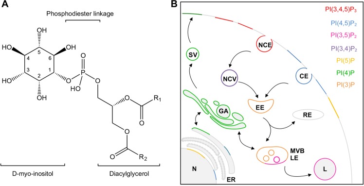
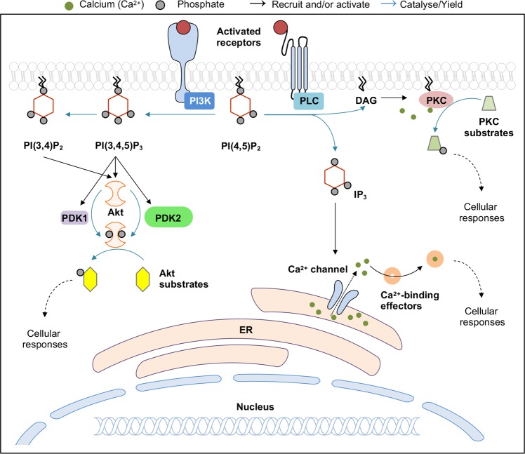
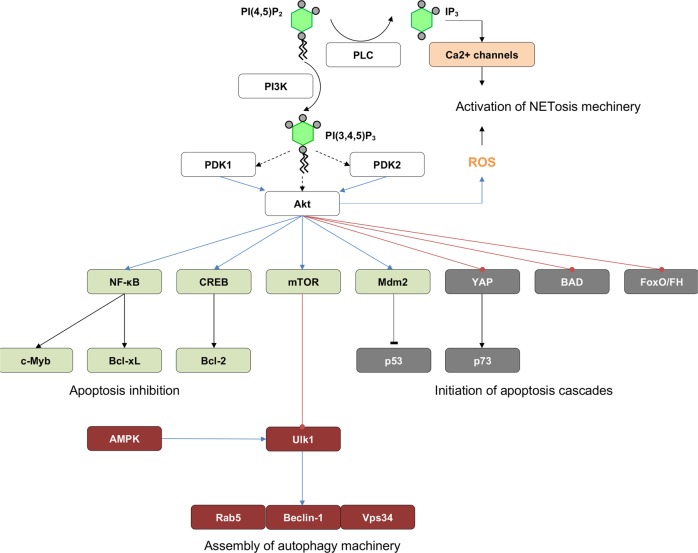
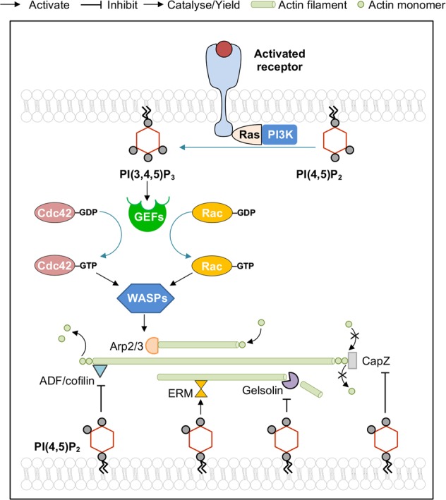
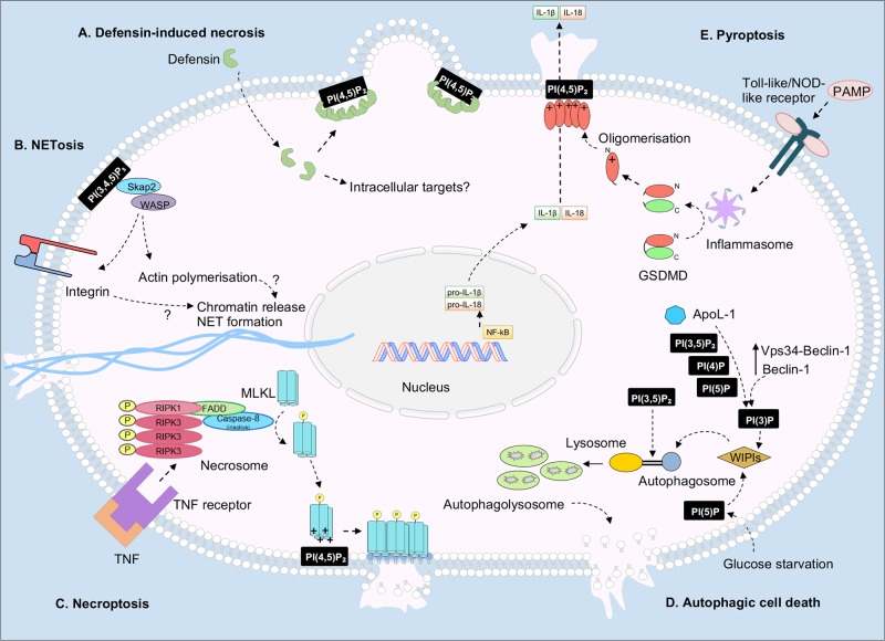
Phosphorylated phosphatidylinositol lipids, or phosphoinositides, critically regulate diverse cellular processes, including signalling transduction, cytoskeletal reorganisation, membrane dynamics and cellular trafficking. However, phosphoinositides have been inadequately investigated in the context of cell death, where they are mainly regarded as signalling secondary messengers. However, recent studies have begun to highlight the importance of phosphoinositides in facilitating cell death execution. Here, we cover the latest phosphoinositide research with a particular focus on phosphoinositides in the mechanisms of cell death. This progress article also raises key questions regarding the poorly defined role of phosphoinositides, particularly during membrane-associated events in cell death such as apoptosis and secondary necrosis. The review then further discusses important future directions for the phosphoinositide field, including therapeutically targeting phosphoinositides to modulate cell death.


Conflict of interest statement

The authors declare that they have no conflict of interest.

Figures

Fig. 1
Fig. 1
Structure and cellular distribution of phosphoinositides. a Chemical structure of phosphatidylinositol, comprising phosphodiester-linked diacylglycerol and D-myo-inositol moieties. R1 and R2 represent any acyl (fatty acid) chain. Hydroxyl at positions 3, 4 and 5 of D-myo-inositol ring are readily phosphorylated, giving rise to seven phosphoinositides. b Localisation of phosphoinositides on plasma and organelle membrane. This distribution map is exemplary, only showing the cellular location where a particular phosphoinositide species is prominently found. CE clathrin-dependent endocytosis, NCE clathrin-independent endocytosis, NCV non-clathrin endocytic vesicles, EE early endosomes, RE recycling endosomes, MVB multivesicular bodies, LE late endosomes, L lysosome, ER endoplasmic reticulum, N nucleus, GA Golgi apparatus, SV secretory vesicles. Black arrow (→) indicates the progression of membrane trafficking pathway
Fig. 2
Fig. 2
Phosphoinositides as a second messengers. PI(4,5)P2 mediates two major transmembrane signalling pathways: PLC-DAG-IP3 and PI3K-Akt. Activated receptor recruits and induce class I PI3K to phosphorylate PI(4,5)P2. The resultant PI(3,4,5)P3 then recruits Akt and its activator kinases PDK1 and PDK2 (or mTORC2, ILK) can also to the plasma membrane, leading to complete phosphorylation of Akt. Alternatively, Akt can also be non-canonically recruited by PI(3,4)P2. Subsequently, Akt regulates downstream effectors through its activated kinase activity. PLC, on the other hand, cleaves PI(4,5)P2 into DAG and IP3 upon stimulation. DAG remains at the plasma membrane to recruit and activate PKC to modulate its downstream effectors via its regulatory phosphorylation. IP3 translocates to ER to excite calcium channels, causing calcium flux into cytoplasm, initiating calcium signalling via calcium binding effectors. PI3K phosphoinositide 3-kinases, PLC phosphoinositide-specific phospholipase C, DAG diacylglycerol, IP3 inositol 1,4,5-trisphosphate, PKC protein kinase C, Akt protein kinase B, PDK1 phosphoinositide-dependent kinase 1, PDK2 phosphoinositide-independent kinase 2, ER endoplasmic reticulum, mTORC2 mechanistic target of rapamycin complex 2, ILK integrin-linked kinase (ILK)
Fig. 3
Fig. 3
Phosphoinositide-mediated signalling pathways in cell death and survival. Proteins involved in (pro-)apoptosis are in dark grey box, anti-apoptosis and survival in green box, autophagy in maroon box and NETosis in orange box. Legends for arrows/lines: kinase-catalysed PIP synthesis (curl arrow), PIP-mediated membrane recruitment (dashed arrow), direct inhibition (blunt-ended solid line), direct activation (solid arrow), stimulatory phosphorylation (blue arrow), inhibitory phosphorylation (red arrow)
Fig. 4
Fig. 4
Regulation of actin remodelling by phosphoinositides PI(3,4,5)P2, synthesised from PI(4,5)P2 upon receptor-mediated stimulation of PI3K via Ras GTPase, recruits GEFs to plasma membrane. GEFs then facilitate GTP loading on Rho family GTPases Cdc42 and Rac, leading to activation of actin nucleators WASPs and, subsequently, Arp2/3 complex. PI(4,5)P2 directly binds to and activates protein mediating membrane-cytoskeleton interaction (e.g. ERM proteins) or inhibits actin depolymerising factor ADF/cofilin, capping proteins (e.g. CapZ) and severing proteins (e.g. gelsolin). PI3K phosphoinositide 3-kinases, Ras Rat sarcoma GTPase, Cdc42 cell division control protein 42 homologue, Rac Ras-related C3 botulinum toxin substrate, GEF guanine nucleotide exchange factor, WASP Wiskott-Aldrich syndrome protein, Arp2/3 actin-related proteins 2/3 complex, ERM erzin, radixin and moesin, ADF actin depolymerising factor
Fig. 5
Fig. 5
Emerging roles of phosphoinositides in cell death. a Defensin-induced necrosis. Defensins internalise into target cells, then bind to PI(4,5)P2 and oligomerise, leading to bleb-associated membrane permeabilisation and cell lysis. b Suicidal NETosis. PI(3,4,5)P3 binding of Src kinase-associated phosphoprotein-2 (Skap2) liberates its active conformation to recruit Wiskott-Aldrich syndrome protein (WASP) for activation of integrins and actin polymerisation, which triggers expulsion of neutrophil extracellular traps (NETs). c Necroptosis. Tumour necrosis factor (TNF) binds to TNF receptor to trigger the formation of the necrosome multiplex, which phosphorylates MLKL and induces its conformational change to expose a PI(4,5)P2-binding domain. Upon PI(4,5)P2 binding, MLKL is recruited to membrane and forms lytic pore to induce membrane rupture. d Autophagic cell death. Lethal autophagy, or autophagic cell death (ACD), occurs upon autophagy overactivation, glucose starvation or death-specific inducer (e.g. lipid-binding ApoL-1). Activation of autophagy effectors (e.g. WIPIs) via PI(3)P- or PI(5)P-mediated pathway leads to the formation of autophagosomes, which are fused with lysosomes, possibly via PI(3,5)P2-dependent manner to form autophagolysomes. Membrane rupture and accumulation of autophagolysosomes are some typical ACD characteristics. e Pyroptosis. Binding of pathogen-associated molecular patterns (PAMPs) to receptors activate the inflammasome to cleave the N-terminus of GSDMD (GSDMD-Nter) from its C-terminus. Through interaction with PI(4,5)P2, GSDMD-NT relocates to the plasma membrane and oligomerises to form lytic membrane pores, causing the release of interleukin 18 (IL-18) and IL-1b

References

    1. Di Paolo G, De Camilli P. Phosphoinositides in cell regulation and membrane dynamics. Nature. 2006;443:651–7. - PubMed
    1. Dickson EJ, Jensen JB, Hille B. Golgi and plasma membrane pools of PI(4)P contribute to plasma membrane PI(4,5)P2 and maintenance of KCNQ2/3 ion channel current. Proc Natl Acad Sci USA. 2014;111:E2281–90. - PMC - PubMed
    1. Sbrissa D, Ikonomov OC, Fu Z, Ijuin T, Gruenberg J, Takenawa T, et al. Core protein machinery for mammalian phosphatidylinositol 3,5-bisphosphate synthesis and turnover that regulates the progression of endosomal transport. Novel Sac phosphatase joins the ArPIKfyve-PIKfyve complex. J Biol Chem. 2007;282:23878–91. - PubMed
    1. Cullen PJ, Carlton JG. Phosphoinositides in the mammalian endo-lysosomal network. Subcell Biochem. 2012;59:65–110. - PMC - PubMed
    1. Sarkes D, Rameh LE. A novel HPLC-based approach makes possible the spatial characterization of cellular PtdIns5P and other phosphoinositides. Biochem J. 2010;428:375–84. - PMC - PubMed

Substances