. 2019 Aug;61(2):244-256.
doi: 10.1165/rcmb.2018-0207OC.

Prolonged Cold Ischemia Induces Necroptotic Cell Death in Ischemia-Reperfusion Injury and Contributes to Primary Graft Dysfunction after Lung Transplantation

Affiliations

Prolonged Cold Ischemia Induces Necroptotic Cell Death in Ischemia-Reperfusion Injury and Contributes to Primary Graft Dysfunction after Lung Transplantation

Xingan Wang et al. Am J Respir Cell Mol Biol. 2019 Aug.

Abstract

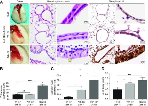
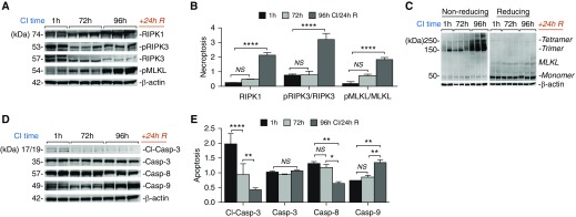
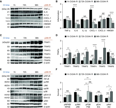
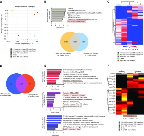
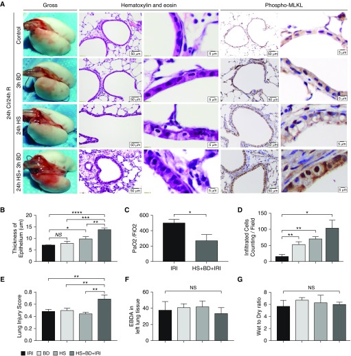
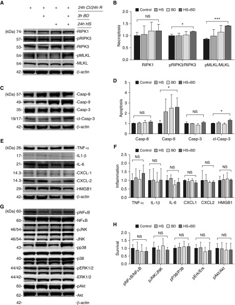
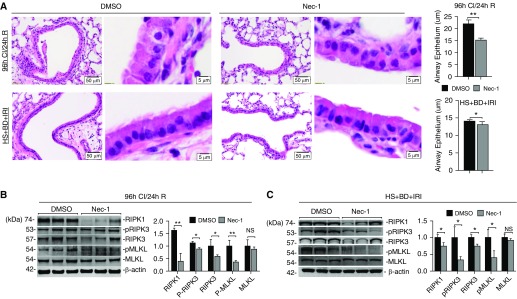
Primary graft dysfunction (PGD) is a major cause of morbidity and mortality after lung transplantation. Ischemia-reperfusion injury (IRI) is a key event that contributes to PGD, though complex interactions affect donor lungs status, such as preceding brain death (BD), hemorrhagic shock (HS), and pre-engraftment lung management, the latter recognized as important risk factors for PGD. We hypothesized that a multi-hit isogenic mouse model of lung transplantation is more closely linked to PGD than IRI alone. Left lung transplants were performed between inbred C57BL/6 mice. A one-hit model of IRI was established by inducing cold ischemia (CI) of the donor lungs at 0°C for 1, 72, or 96 hours before engraftment. Multi-hit models were established by inducing 24 hours of HS and/or 3 hours of BD before 24 hours of CI. The recipients were killed at 24 hours after transplant and lung graft samples were analyzed. In the one-hit model of IRI, up to 72-hour CI time resulted in minimal cellular infiltration near small arteries after 24-hour reperfusion. Extension of CI time to 96 hours led to increased cellular infiltration and necroptotic pathway activation, without evidence of apoptosis, after 24-hour reperfusion. In a multi-hit model of PGD, "HS + BD + IRI" demonstrated increased lung injury, cellular infiltration, and activation of necroptotic and apoptotic pathways compared with IRI alone. Treatment with an inhibitor of receptor-interacting protein kinase 1 kinase, necrostatin-1, resulted in a significant decrease of downstream necroptotic pathway activation in both single- and multi-hit models of IRI. Thus, activation of necroptosis is a central event in IRI after prolonged CI, though it may not be sufficient to cause PGD alone. Pathological evaluation of donor lungs after CI-induced IRI, in conjunction with pre-engraftment donor lung factors in our multi-hit model, demonstrated early evidence of lung injury consistent with PGD. Our findings support the premise that pre-existing donor lung status is more important than CI time alone for inflammatory pathway activation in PGD, which may have important clinical implications for donor lung retrieval.

Keywords: lung transplantation; necroptosis; primary graft dysfunction; reperfusion injury.


Figures

Figure 1.
Figure 1.
Prolonged cold ischemia (CI) worsens ischemia–reperfusion injury in a mouse model of orthotopic lung transplantation. Using murine syngeneic lung transplantation between C57BL/6 mice (n = 3 mice per group), cold preservation time was prolonged up to 96 hours at 0°C to induce varying severities of CI/repurfusion (R) (A). Shown in A are changes in gross appearance of lungs; low (scale bars = 5 μm) and high magnification (scale bars = 50 μm) of the lung alveolar capillary units. Shown after 96-hour CI, the epithelial cells of the grafted lung manifested swelling with increased translucency, but the nuclei remained intact (A, yellow arrow) using hematoxylin and eosin staining. Phospho-mixed-lineage kinase domain-like kinase (MLKL) staining (right panels) is predominantly localized to the airway epithelium and is markedly increased in the 96-hour CI/24-hour R group compared with the earlier time points. (B) The thickness of the epithelial barrier was measured, demonstrating increased cross-sectional area after 96 hours of CI time. Lung injury scores were determined by evaluating neutrophils in the alveolar and interstitial space, the presence of hyaline membranes, airspace proteinaceous debris, and alveolar septal thickening (22). (C and D) Prolonged CI led to a significant increase of infiltrating inflammatory cells (C) and lung injury (D) of the grafts. Student’s t test. *P < 0.05, **P < 0.01, ***P < 0.001, and ****P < 0.0001. A two-way ANOVA was performed for statistical analysis. Data are expressed as mean (±SEM). NS = not significant.
Figure 2.
Figure 2.
CI–reperfusion injury activates necroptosis after experimental orthotopic lung transplantation. (A) Immunoblot analysis showing levels of necroptosis effectors receptor-interacting protein kinase (RIPK) 1, pRIPK3, and phospho (p) MLKL are increased after CI–reperfusion injury. (B) Densitometric analysis of immunoblots show significantly increased RIPK1 after normalization to actin (arbitrary units), and ratio of pMLKL/MLKL, pRIPK3/RIPK3 after prolonged CI. (C) As a consequence of MLKL phosphorylation, we observed the formation of MLKL trimer/tetramer complexes consistent with necrosome assembly after 96-hour CI and reperfusion. (D) Levels of death effector caspases are shown by immunoblotting. Cleaved caspase-3 and caspase-8 decreased significantly in the grafted lung after 96-hour CI and reperfusion. (E) Densitometric analysis of immunoblots in D after normalization to actin showing loss of caspase-8, a necroptosis inhibitor, and capase-3, an effector of apoptosis, as the CI time was extended. Each lane in the immunoblots represents protein isolated from a separate mouse whole lung allograft, CI time; n = 2 at 1 hour, n = 3 at 72 hours and 96 hours. A two-way ANOVA was used for statistical analysis. *P < 0.05, **P < 0.01, and ****P < 0.0001. Data are expressed as mean (±SEM).
Figure 3.
Figure 3.
CI–reperfusion injury activates inflammatory signaling after experimental orthotopic lung transplantation. (A) Immunoblot showing unchanged or decreased levels of several proinflammatory cytokines and the damage-associated molecular pattern, HMGB1 (high mobility group box 1). (B) Densitometric analysis after actin normalization of immunoblots in A showing significantly decreased levels of IL-6, CXCL-1, and HMGB1 during CI and R. (C) Levels of TNF-associated factor (TRAF) proteins, which are involved in necroptosis pathway activation, are shown by immunoblotting. (D) Densitometric analysis of immunoblots in C showing that TRAFs (except TRAF6) markedly increased in the grafted lung tissue as CI time was extended to 96 hours. (E and F) Various cell stress and survival kinases were assayed by immunoblot in E and densitometry of bands quantitated as a ratio of phosphorylated to total protein in F. Each lane in the immunoblots represents protein isolated from a separate mouse whole-lung allograft over CI time; n = 2 at 1 hour, n = 3 at 72 hours and 96 hours. A two-way ANOVA was used for statistical analysis. *P < 0.05, **P < 0.01, and ****P < 0.0001. Data are expressed as mean (±SEM).
Figure 4.
Figure 4.
Comparative transcriptomic analysis of the effect of prolonged donor lung CI in experimental lung transplantation. A basic transcriptomic analysis of donor lungs before and after engraftment with reperfusion by RNA sequencing (n = 1 per treatment group). (A) Principal component analysis scatter plot revealing covariance of the 24- and 96-hour donor lung CI, which indicates close similarity in the gene expression profile. After 2 hours of reperfusion, there is a distinct alteration of the projection of the 48- and 96-hour transcriptome compared with control lung reperfusion. (B) Venn diagram representation of differentially expressed genes (DEGs) from 48- and 96-hour donor lung CI compared with control donor lung; 3,544 genes and 2,995 genes, respectively, demonstrated significant overlap of 1,414 genes between the groups; representation factor 3.3, P < 0.0001. Reported genes were filtered with a minimum absolute fold change of 1.5 and a false discovery rate (FDR) P < 0.05. Pathway analysis of genes that were common to both groups revealed processes affecting cell cycle progression, IL-6 signaling, and extracellular matrix (ECM) interactions. (C) Hierarchical cluster heatmap representation of the top 500 DEGs demonstrating clustering of control donor lungs and those subjected to 24- and 96-hour CI compared with a divergent gene expression profile after 2 hours of reperfusion. (D) There is significant overlap of 3,197 genes that are differentially expressed after 2 hours of reperfusion in donor lungs subjected to 24 hours (3,498 DEGs) and 96 hours (4,098 DEGs) of CI time; representation factor 5.6; P < 0.0001. (E) Common and unique DEGs from 24-hour and 96-hour CI after 2 hours of reperfusion, as visualized in the Venn diagram in D, were subjected to pathway analysis using statistical overrepresentation testing in Panther with Benjamini and Hochberg FDR correction. Reperfusion resulted in significant enrichment of genes that involve pathway activation of complement C3 and C5, necroptotic cell death, and inflammatory IL-6 signaling. There was selective enrichment of genes involved in respiratory electron transport in the donor lung after 24-hour CI, and the cellular response to stress in the donor lung after 96-hour CI, after 2 hours of reperfusion, respectively. (F) Heatmap visualization and hierarchical clustering of a necroptotic pathway gene set identified during pathway analysis.
Figure 5.
Figure 5.
Pathological characteristics of primary graft dysfunction (PGD) are demonstrated in a clinically relevant multi-hit model of lung injury. To mimic clinical settings, pre-existing donor lung injury was induced by subjecting donor mice to hemorrhagic shock (HS) and/or brain death (BD) in addition to 24-hour CI time (n = 3 mice per group). (A) The three-hit model demonstrates more significant hyaline membrane deposition, bronchial epithelial injury, and inflammatory infiltration around small arteries. Shown are changes in gross appearance of lungs, and low (scale bars = 50 μm) and high magnification (scale bars = 5 μm) of the lung alveolar capillary units. In the three-hit model of HS + BD + ischemia–reperfusion injury (IRI), severity of lung injury was most prominent compared with either one-hit or two-hit models using hematoxylin and eosin staining. pMLKL staining is predominantly localized to the airway epithelium and is increased in the HS + BD + IRI multi-hit model compared with the single-hit models of IRI. (B) Epithelial thickness was increased in the three-hit model compared with control and single-hit models. (C) The ratio of PaO2 to FiO2 showed that graft function remained normal in the one-hit model of IRI but deteriorated significantly in three-hit model of HS + BD + IRI. There was significantly more inflammatory infiltration (D) and higher acute lung injury scores (E) in the three-hit model of HS + BD + IRI. (F and G) The test of Evans-blue dye albumin (EBDA) leakage and wet-to-dry ratio showed no difference in permeability of microvasculature between one-hit and multi-hit models. *P < 0.05, **P < 0.01, ***P < 0.001, and ****P < 0.0001. Two-way ANOVA testing was used for statistical analysis. Data are expressed as mean (±SEM).
Figure 6.
Figure 6.
Necroptosis contributes to regulated cell death in a multi-hit model of PGD. Immunoblotting analysis showing effector levels of necroptosis, apoptosis, inflammation, and signaling kinases (n = 3 mice per group). (A) The phosphorylation status of MLKL increased with pre-existing donor lung injury. (B) Densitometric analysis after actin normalization of immunoblots in A showing significantly increased ratios of phosphorylated MLKL (pMLKL) to total MLKL and of pRIPK3 to total RIPK3. (C) In contrast to repressed apoptosis in prolonged CI–reperfusion injury, cleaved caspase-3 increased in the three-hit model of HS + BD + IRI. (D) Densitometric analysis revealed a degree of intrinsic apoptotic pathway activation in addition to necroptosis, which differentiates the mechanisms of cell death activation in the multi-hit model from prolonged cold IRI alone. (EH) There was no difference detected in levels of proinflammatory cytokines or signaling kinases in lung grafts with or without pre-existing injury. *P < 0.05 and ***P < 0.001. Two-way ANOVA testing was used for statistical analysis. Data are expressed as mean (±SEM).
Figure 7.
Figure 7.
Necrostatin attenuates necroptotic cell death activation after lung transplantation. Recipient mice were administered DMSO (control) or necrostatin (Nec)-1 (10 mg/kg) 2 hours before lung transplantation (n = 3 mice per group). (A) Pathologic changes of the lung airway epithelium at low (scale bars = 50 μm) and high magnification (scale bars = 5 μm) after treatment with Nec-1 or DMSO. Nec-1 treatment resulted in a significant reduction in airway epithelial thickness in the 96-hour CI/24-hour R group and a small, but statistically significant, reduction in airway epithelial thickness in the HS + BD + IRI group; **P < 0.01 and *P < 0.05, respectively, using a two-tailed Student’s t test. Corresponding immunoblot analysis shows levels of RIPK1 and its downstream effectors are decreased significantly after the treatment of Nec-1 in both one-hit model of prolonged cold IRI (B) and three-hit model of HS + BD + IRI (C); *P < 0.05 and **P < 0.01 versus DMSO using two-way ANOVA. Data are expressed as mean (±SEM).

Comment in

Similar articles

Cited by

References

    1. Diamond JM, Arcasoy S, Kennedy CC, Eberlein M, Singer JP, Patterson GM, et al. Report of the International Society for Heart and Lung Transplantation Working Group on Primary Lung Graft Dysfunction, part II: epidemiology, risk factors, and outcomes—a 2016 Consensus Group statement of the International Society for Heart and Lung Transplantation. J Heart Lung Transplant. 2017;36:1104–1113. - PubMed
    1. Suzuki Y, Cantu E, Christie JD. Primary graft dysfunction. Semin Respir Crit Care Med. 2013;34:305–319. - PMC - PubMed
    1. Gelman AE, Fisher AJ, Huang HJ, Baz MA, Shaver CM, Egan TM, et al. Report of the ISHLT Working Group on Primary Lung Graft Dysfunction part III: mechanisms. A 2016 Consensus Group statement of the International Society for Heart and Lung Transplantation. J Heart Lung Transplant. 2017;36:1114–1120. - PMC - PubMed
    1. Fiser SM, Tribble CG, Long SM, Kaza AK, Kern JA, Jones DR, et al. Ischemia–reperfusion injury after lung transplantation increases risk of late bronchiolitis obliterans syndrome. Ann Thorac Surg. 2002;73:1041–1047. [Discussion, pp. 1047–1048.] - PubMed
    1. Whitson BA, Prekker ME, Herrington CS, Whelan TP, Radosevich DM, Hertz MI, et al. Primary graft dysfunction and long-term pulmonary function after lung transplantation. J Heart Lung Transplant. 2007;26:1004–1011. - PubMed

Publication types

MeSH terms