. 2019 Jul;61(1):42-50.
doi: 10.1165/rcmb.2018-0236MA.

A Flow Cytometric Method for Isolating Cystic Fibrosis Airway Macrophages from Expectorated Sputum

Affiliations

A Flow Cytometric Method for Isolating Cystic Fibrosis Airway Macrophages from Expectorated Sputum

Katherine B Hisert et al. Am J Respir Cell Mol Biol. 2019 Jul.

Abstract

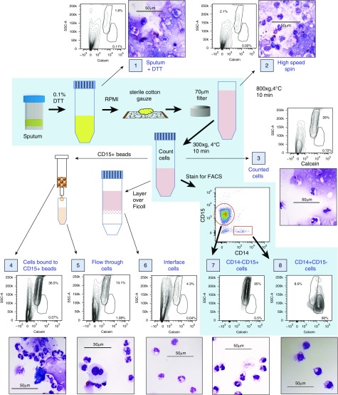
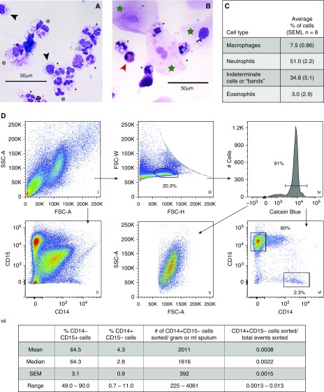
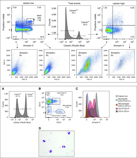
Research to understand the contribution of macrophages to nonresolving airway inflammation in cystic fibrosis (CF) and other chronic suppurative airways diseases has been hindered by a lack of methods for isolating and studying these cells. With the development of technologies that can characterize small numbers of cells or individual cells, there is an even greater need for methodologies to isolate rare cells in heterogeneous specimens. Here, we describe a method that overcomes the technical obstacles imposed by sputum debris and apoptotic cells, and allows isolation of pure populations of macrophages from CF sputum. In addition to enhancing our ability to study human CF airway macrophages, this protocol can be adapted to study cells in sputum from other chronic suppurative lung diseases (e.g., chronic obstructive pulmonary disease) and used for isolation of individual cells for single cell analyses.

Keywords: cystic fibrosis; macrophages; single-cell analyses; sputum.


Figures

Figure 1.
Figure 1.
Efficacy of different methods for isolating leukocytes from spontaneously expectorated cystic fibrosis (CF) sputum evaluated by flow cytometry and histologic examination. Steps in the protocol described in this article are depicted on the blue background, with thick black arrows between each step. Samples of a single specimen were evaluated at steps throughout the protocol (each sample is identified by a number in a blue box) to determine the presence of debris and the purity of the leukocyte populations by both flow cytometry and histologic examination. Thin black arrows indicate samples produced either by initial steps in the protocol or by other protocols that are commonly used to isolate leukocyte populations but are ineffective for isolating sputum leukocytes. Side scatter-area (SSC-A) versus calcein (a viability dye that fluoresces only when taken up by intact, viable cells) can be used to separate live cells and debris, and can distinguish neutrophils from macrophages more effectively than forward scatter-area (FSC-A). Larger images of histology for each sample can be found in Figure E2. Scale bars: 50 μm.
Figure 2.
Figure 2.
CF sputum contains macrophages and neutrophils that can be sorted as pure populations by FACS. (A and B) Morphologic identification of sputum leukocytes. Sputum specimens were solubilized with DTT, strained, filtered, and centrifuged before counting (as in Figure E1), and ∼30,000 leukocytes were applied to a microscope slide via cytospin and stained with Romanowsky stain. Two separate specimens are shown. Macrophages (black arrowheads) have purple-blue cytoplasm lacking granules, and a single, eccentric nucleus. Neutrophils (black asterisks) have pink cytoplasm containing granules, and a multilobular nucleus. Eosinophils (red arrowhead) are binucleate with red cytoplasmic granules. Indeterminate cells (at symbols) have a single nucleus, and pink and/or granular cytoplasm; these were determined later to be neutrophils by flow cytometry. Squamous cells are large, with a “fried egg” appearance (green star). (C) Quantitation of cell differentials from several sputum specimens as determined by light microscopy (n = 8, collected from three subjects). (D) Flow cytometric parameters for sorting macrophages and neutrophils from solubilized sputum specimens. (i, ii, and iii) Total events plotted for the indicated parameters. (iii) Gating strategy for single cells. (iv) Plot of single cells, gating on calcein+ cells. (v) Live single cells (identified in iv) plotted for FSC-A versus SSC-A (compare with i). (vi) Live single cells plotted for CD14 versus CD15 (compare with ii). (vii) Percentage of different cell subsets in sputum specimens from different subjects, as determined by flow cytometry, as well as the average numbers of macrophages per gram or milliliter of sputum that can be sorted using this method. n = 17 sputum specimens from 10 different subjects; repeat specimens (from subjects 3 and 18) were obtained on multiple days over several months. >50,000 neutrophils were sorted from virtually all sputum specimens, including those < 1.0 gram in weight. FSC-H = forward scatter-height; FSC-W = forward scatter-width. Scale bars: 50 μm.
Figure 3.
Figure 3.
Flow cytometric analysis of specimens stained with propidium iodide (PI) and annexin-V reveals apoptotic neutrophils in CF sputum. PI and annexin-V staining was used to identify apoptotic cell populations in CF sputum cells. PI binds to DNA in cells in which membrane integrity has been lost (dead cells), and annexin-V binds to accessible phosphatidylserine in apoptotic or dead cells. Thus, annexin+/PI cells are apoptotic, and annexin+/PI+ cells are necrotic. Annexin/PI events are either debris (calcein) or viable cells (calcein+). Top and bottom panels display data from different sputum specimens. Top: Total events were separated into calceinhi (live) and calceinlow (dead/debris) populations, and then plotted for annexin-V versus PI. All PI cells were then plotted for FSC-A versus SSC-A. As expected, the majority of live cells (calceinhi) were comprised of nonapoptotic cells. The calceinlow events contained a majority of apoptotic and dead cells. Bottom: (A) In a different sample, total events were plotted to determine calcein positivity. (B) Viable cells (calceinhi) were then plotted for CD14 and CD15 expression, and four populations were defined (polymorphonuclear cells [PMN] or CD15hiCD14, CD15hiCD14lo, CD15CD14, and macrophages or CD15CD14+). (C) Annexin-V binding to the four populations delineated in B, plus the calceinlow cells in A; CD15/CD14 cells in the black histogram are behind the macrophage histogram depicted in orange. (D) Cytospins of sorted apoptotic cells (calcein+/annexin+/PI) reveal that these cells are neutrophils.

References

    1. Gibson RL, Burns JL, Ramsey BW. Pathophysiology and management of pulmonary infections in cystic fibrosis. Am J Respir Crit Care Med. 2003;168:918–951. - PubMed
    1. Mogayzel PJ, Jr, Naureckas ET, Robinson KA, Mueller G, Hadjiliadis D, Hoag JB, et al. Pulmonary Clinical Practice Guidelines Committee. Cystic fibrosis pulmonary guidelines: chronic medications for maintenance of lung health. Am J Respir Crit Care Med. 2013;187:680–689. - PubMed
    1. Johnston LK, Rims CR, Gill SE, McGuire JK, Manicone AM. Pulmonary macrophage subpopulations in the induction and resolution of acute lung injury. Am J Respir Cell Mol Biol. 2012;47:417–426. - PMC - PubMed
    1. Murray PJ, Allen JE, Biswas SK, Fisher EA, Gilroy DW, Goerdt S, et al. Macrophage activation and polarization: nomenclature and experimental guidelines. Immunity. 2014;41:14–20. - PMC - PubMed
    1. Parisi L, Gini E, Baci D, Tremolati M, Fanuli M, Bassani B, et al. Macrophage polarization in chronic inflammatory diseases: killers or builders? J Immunol Res. 2018;2018:8917804. - PMC - PubMed

Publication types