. 2019 Feb 11;13(2):e0007080.
doi: 10.1371/journal.pntd.0007080. eCollection 2019 Feb.

Detection of Zika virus in mouse mammary gland and breast milk

Affiliations

Detection of Zika virus in mouse mammary gland and breast milk

Jose Angel Regla-Nava et al. PLoS Negl Trop Dis. .

Abstract

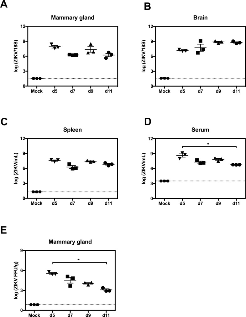
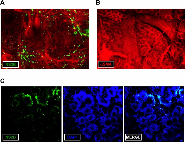
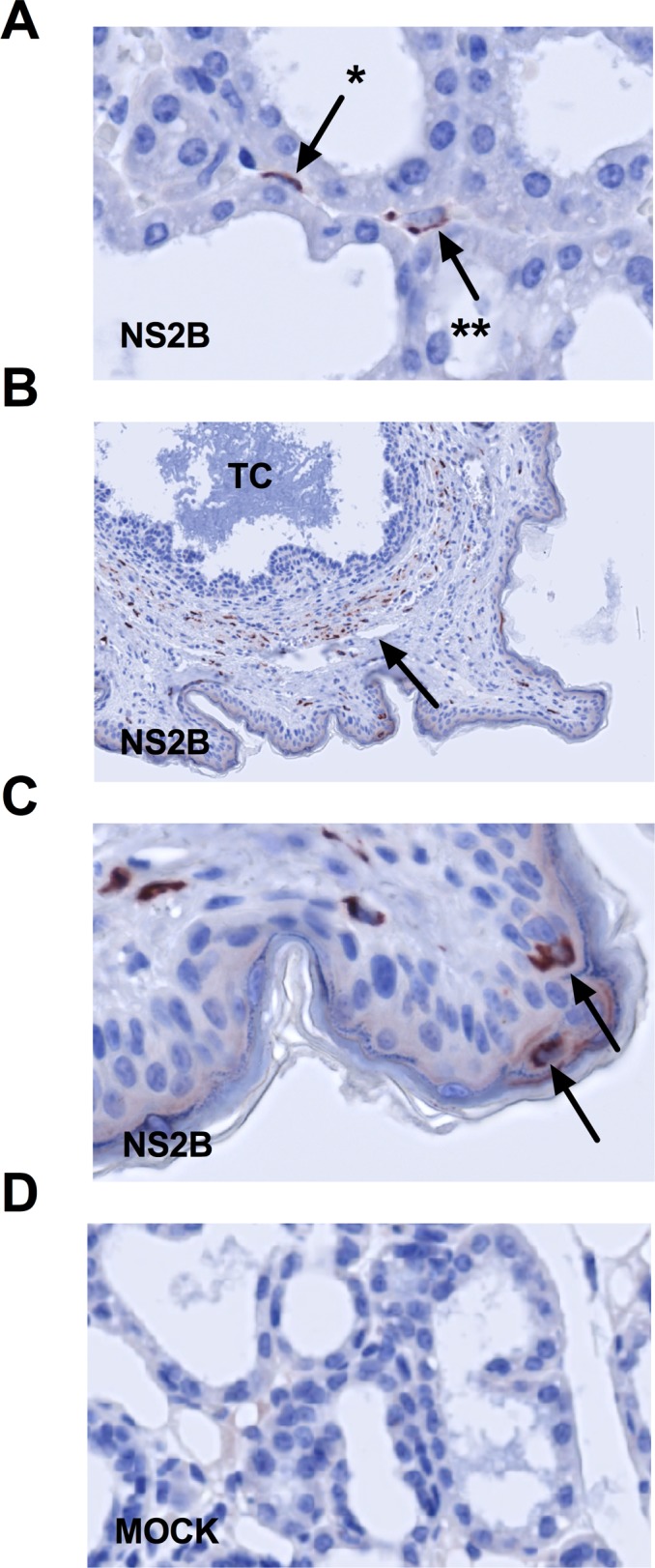
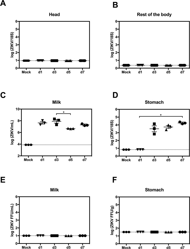
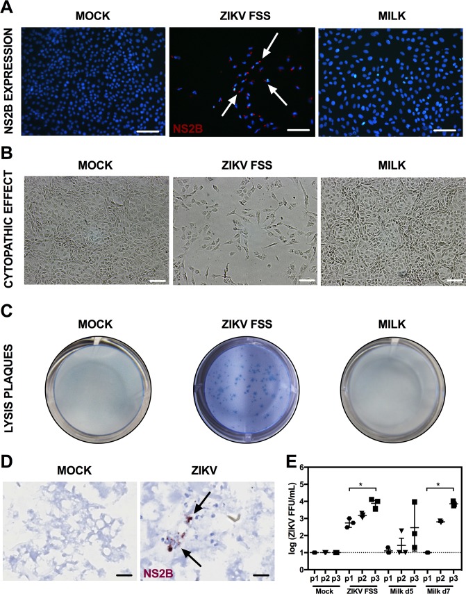
Clinical reports of Zika Virus (ZIKV) RNA detection in breast milk have been described, but evidence conflicts as to whether this RNA represents infectious virus. We infected post-parturient AG129 murine dams deficient in type I and II interferon receptors with ZIKV. ZIKV RNA was detected in pup stomach milk clots (SMC) as early as 1 day post maternal infection (dpi) and persisted as late as 7 dpi. In mammary tissues, ZIKV replication was demonstrated by immunohistochemistry in multiple cell types including cells morphologically consistent with myoepithelial cells. No mastitis was seen histopathologically. In the SMC and tissues of the nursing pups, no infectious virus was detected via focus forming assay. However, serial passages of fresh milk supernatant yielded infectious virus, and immunohistochemistry showed ZIKV replication protein associated with degraded cells in SMC. These results suggest that breast milk may contain infectious ZIKV. However, breast milk transmission (BMT) does not occur in this mouse strain that is highly sensitive to ZIKV infection. These results suggest a low risk for breast milk transmission of ZIKV, and provide a platform for investigating ZIKV entry into milk and mechanisms which may prevent or permit BMT.


Conflict of interest statement

M.J. is CEO of Visikol.

Figures

Fig 1
Fig 1. ZIKV replication in mammary glands of AG129 mice.
Postpartum 8-week-old AG129 dams were retro-orbitally inoculated with 1 x 10² FFU of ZIKV FSS13025 or 10% FBS-PBS as Mock within 24 hours of parturition. (A-D) Viral titers in mammary gland, brain, spleen and serum were determined via qRT-PCR at 5, 7, 9 and 11 days post infection (dpi). (E) Levels of infectious ZIKV in mammary gland were determined by FFA at 5, 7, 9 and 11 dpi. Negative controls were evaluated at 5 days after mock-infection (A-E). n = 3 mice per time point in each panel. Data represent two independent experiments.
Fig 2
Fig 2. Max intensity z-projection of ZIKV-infected mammary tissue.
Postpartum AG129 dams were inoculated via retro-orbital route with 1 x 10² FFU of ZIKV FSS13025 or 10% FBS-PBS as Mock within 24 hours of parturition and sacrificed at 5 dpi. (A) Staining of mammary gland from ZIKV-infected mice with anti-αSMA (red) and anti-ZIKV NS2B antibodies (green). (B) Staining of mammary tissue from mock-infected mice with anti-αSMA (red) and anti-ZIKV NS2B antibodies (green). (C) Staining of mammary tissue from ZIKV-infected mice with anti-ZIKV NS2B (green) and DAPI (blue). Tissues were imaged in sequential mode at 2x digital zoom on a 10x air objective (20x overall). Representative data from 3 ZIKV-infected and 3 mock-infected mice are shown.
Fig 3
Fig 3. Immunohistochemical detection of ZIKV in mammary gland of AG129 mice.
Postpartum AG129 dams were retro-orbitally inoculated with 1 x 10² FFU of ZIKV FSS13025 or 10% FBS-PBS as Mock within 24 hours of parturition and sacrificed at 5 and 9 dpi. (A) Mammary gland was removed and ZIKV NS2B immunoreactivity was detected at 5 dpi. ZIKV NS2B immunoreactivity was detected in (*) myoepithelial cells and probable macrophages (**) in the interstitium. (B) At 9 dpi, many cells expressing ZIKV NS2B were seen in cells in the stroma surrounding teat canal. (C) Magnification of teat: There was strong ZIKV NS2B expression in some Langerhans cells. (D) Negative control mammary tissue from a mock-infected mouse is shown. Arrows indicate NS2B-expressing cells. TC, teat canal; n = 3 mice per group.
Fig 4
Fig 4. Transmission of ZIKV RNA in mouse breast milk.
Postpartum AG129 dams were retro-orbitally inoculated with 1 x 10² FFU of ZIKV FSS13025 or 10% FBS-PBS as Mock within 24 hours of parturition. Pups were sacrificed on d1, d3, d5 and d7 after birth. (A and B) Levels of ZIKV RNA in the head and the rest of the body were measured by qRT-PCR (n = 6 mice, each day). (C and D) ZIKV RNA levels in stomach milk clots (SMC) and stomach tissues were quantified via qRT-PCR (n = 3 mice, each day). (E and F) Presence of infectious ZIKV in SMC and stomach was assessed using FFA (n = 3 mice, each day). Data represent two independent experiments.
Fig 5
Fig 5. Infectivity of milk supernatant on vero cells.
AG129 dams were retro-orbitally inoculated with 1 x 10² FFU of ZIKV FSS13025 within 24 hours of parturition. (A-C) Vero cells were mock-infected as a negative control, infected with ZIKV FSS13025 at an MOI of 0.001 as a positive control, cultured with stomach milk clot (SMC) supernatant collected from the pup stomachs 3 days after birth, or (D) cultured with fresh milk obtained directly from dams at days 5 and 7 after infection; mock fresh milk was collected on day 5 after parturition. (A) Vero cells were fixed at day 5 after infection or culture with SMC supernatant. ZIKV NS2B protein (red) was labeled with a specific antibody. Nuclei were stained with DAPI (blue). Arrows indicate cells expressing ZIKV NS2B protein. (B) CPE and (C) plaques were visualized at at day 5 after infection or culture with SMC supernatant. Slides for immunofluorescence were examined on a Nikon/eclipse 80 microscope and CPE on an Eclipse TE300 microscopy and Nikon DXM1200C camera. (D) Postpartum AG129 mice were retro-orbitally inoculated with 1 x 10² FFU of ZIKV FSS13025 or 10% FBS-PBS as mock within 24 hours of parturition. Pups were sacrificed on day 5 after birth. ZIKV NS2B expression was determined in SMC via IHC. Negative control SMC samples are obtained from pups born to mock-infected mice. Representative images from n = 10 mice per group are shown. (E) Levels of infectious ZIKV in the fresh milk samples were determined after serial passages in Vero cells using FFA. The Vero cell culture supernatants were collected at day 3 after culture with fresh milk, and this passaging was repeated for 2 additional rounds.; n = 3 mice per group.

References

    1. Lazear H.M. and Diamond M.S., Zika Virus: New Clinical Syndromes and its Emergence in the Western Hemisphere. J Virol, 2016. - PMC - PubMed
    1. Tang W.W., et al., A Mouse Model of Zika Virus Sexual Transmission and Vaginal Viral Replication. Cell Rep, 2016. 17(12): p. 3091–3098. 10.1016/j.celrep.2016.11.070 - DOI - PMC - PubMed
    1. Newman C.M., et al., Oropharyngeal mucosal transmission of Zika virus in rhesus macaques. Nat Commun, 2017. 8(1): p. 169 10.1038/s41467-017-00246-8 - DOI - PMC - PubMed
    1. Grischott F., et al., Non-vector-borne transmission of Zika virus: A systematic review. Travel Med Infect Dis, 2016. 14(4): p. 313–30. 10.1016/j.tmaid.2016.07.002 - DOI - PubMed
    1. Blohm G.M., et al., Complete Genome Sequences of Identical Zika virus Isolates in a Nursing Mother and Her Infant. Genome Announc, 2017. 5(17). - PMC - PubMed

Publication types

LinkOut - more resources