. 2019 May;111(5):1317-1334.
doi: 10.1111/mmi.14223. Epub 2019 Mar 26.

The master quorum-sensing regulators LuxR/HapR directly interact with the alpha subunit of RNA polymerase to drive transcription activation in Vibrio harveyi and Vibrio cholerae

Affiliations

The master quorum-sensing regulators LuxR/HapR directly interact with the alpha subunit of RNA polymerase to drive transcription activation in Vibrio harveyi and Vibrio cholerae

Alyssa S Ball et al. Mol Microbiol. 2019 May.

Abstract

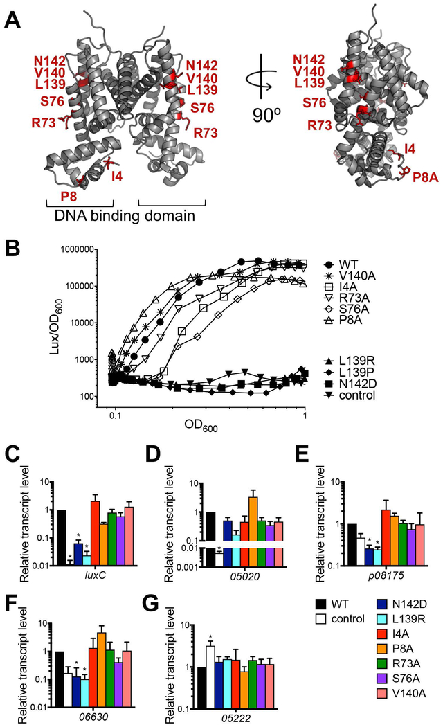
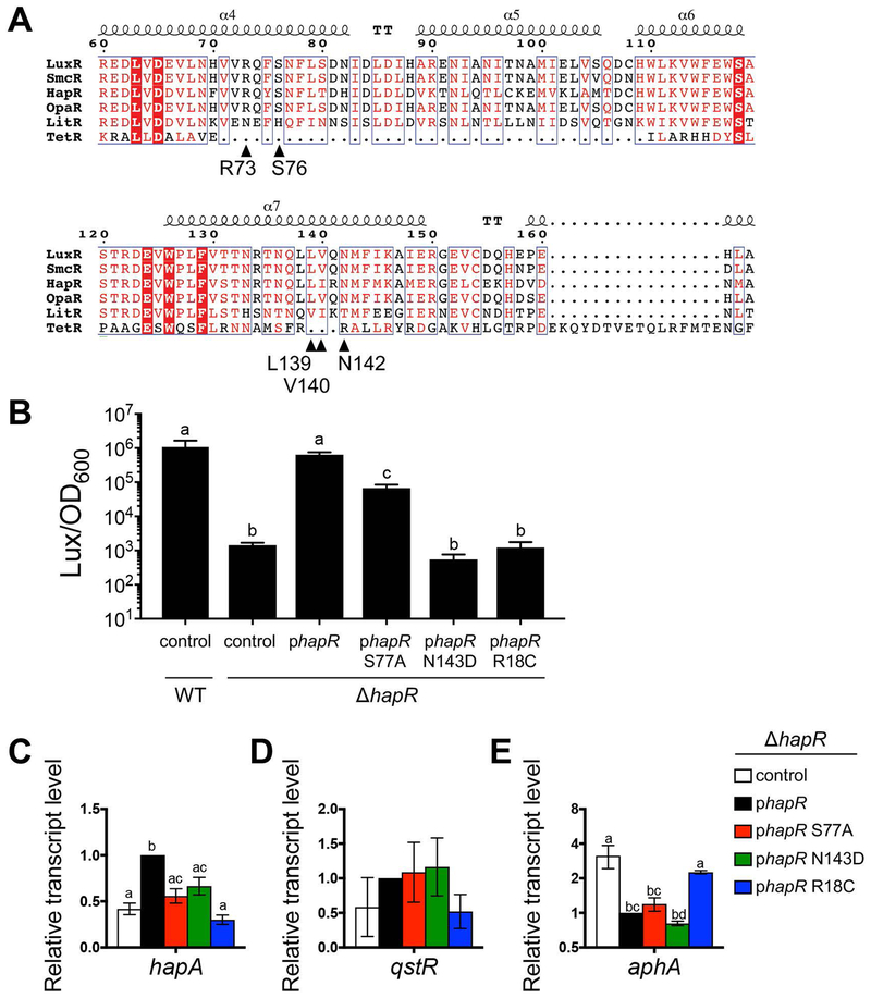
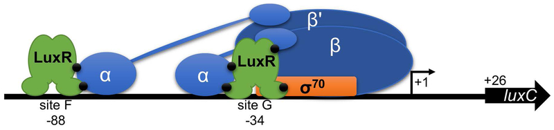
In Vibrio species, quorum sensing controls gene expression for numerous group behaviors, including bioluminescence production, biofilm formation, virulence factor secretion systems, and competence. The LuxR/HapR master quorum-sensing regulators activate expression of hundreds of genes in response to changes in population densities. The mechanism of transcription activation by these TetR-type transcription factors is unknown, though LuxR DNA binding sites that lie in close proximity to the -35 region of the promoter are required for activation at some promoters. Here, we show that Vibrio harveyi LuxR directly interacts with RNA polymerase to activate transcription of the luxCDABE bioluminescence genes. LuxR interacts with RNA polymerase in vitro and in vivo and specifically interacts with both the N- and C-terminal domains of the RNA polymerase α-subunit. Amino acid substitutions in the RNAP interaction domain on LuxR decrease interactions between LuxR and the α-subunit and result in defects in transcription activation of quorum-sensing genes in vivo. The RNAP-LuxR interaction domain is conserved in Vibrio cholerae HapR and is required for activation of the HapR-regulated gene hapA. Our findings support a model in which LuxR/HapR bind proximally to RNA polymerase to drive transcription initiation at a subset of quorum-sensing genes in Vibrio species.


Conflict of interest statement

Conflict of Interest Statement

The authors declare that that they have no conflicts of interest to declare.

Figures

Figure 1.
Figure 1.. LuxR interacts with RNAP in vivo and in vitro.
(A) Elutions from IP reactions with a ΔluxR V. harveyi strain (KM669) containing plasmids expressing either FLAG-LuxR (pAP116), an empty vector control plasmid (pSLS3), or FLAG-R17C (pST012). The bands corresponding to the FLAG-LuxR protein and the light chain antibody to the FLAG epitope are indicated. (B) Western blot analyses of lysates and elutions from IP reactions with a ΔluxR V. harveyi strain (KM669) containing either a plasmid expressing FLAG-LuxR (pAP116) or an empty vector control plasmid (pSLS3). Purified FLAG-LuxR protein (1 μg) was included in lane 1 as a positive control for the FLAG western. For each strain, two IP reactions were tested with differing stringencies in the wash steps: 1) 100 mM NaCl, 0.1% Triton-X, and 2) 500 mM NaCl, 1% Triton-X. Antibodies used are indicated on the left of each panel. (C, D) SDS-PAGE (C) and western blot (D) analyses of co-IP experiments with purified FLAG-LuxR and His-RNAP holoenzyme incubated with anti-FLAG resin. Purified E. coli His-RNAP (216 pmol) and purified FLAG-LuxR (54 pmol) were loaded in lanes 2 and 3, respectively, in panel C. Samples were taken of the supernatants (S) after incubation with the anti-FLAG resin and samples of the elution (E) were collected from the resin after elution. The western blot was probed with antibodies against β’. (E) Western blot analysis of reciprocal co-IP experiments performed with FLAG-LuxR and His-RNAP incubated with nickel-NTA resin. Samples were taken of the supernatants (S) after incubation with the nickel-NTA resin and samples of the elution (E) were collected from the resin after elution. The western blot was probed with antibodies against the FLAG epitope.
Figure 2.
Figure 2.. LuxR DNA binding to PluxC promoter binding sites.
(A) Diagram of the luxCDABE promoter region. Gray boxes indicate LuxR binding sites (BS) and hatched boxes are predicted σ BSs. The primary transcription start site is indicated by a black arrow at +1, and the upstream low cell density transcription start site is indicated by a black arrow at −97. (B, C, D) Electrophoretic mobility shift assays were conducted with dsDNA substrates corresponding to LuxR binding sites B (panel B; oligos JCV617, JCV363), F (panel C; oligos AB190, AB191), or G (panel D; oligos AB188, AB189) with purified proteins LuxR, LuxR N142D, and LuxR L139R at concentrations ranging from 0.5 to 500 nM. The percentage of DNA bound is graphed for each protein. A two-way analysis of variance (ANOVA) followed by Tukey’s multiple-comparison test (n=3) was performed on all data points, and no statistical differences were observed (p < 0.001).
Figure 3.
Figure 3.. LuxR interacts with the N-terminal domain of RNAP α subunit.
(A) Western blot analysis of co-IP experiments with purified FLAG-LuxR and E. coli His-α incubated with anti-FLAG resin. Samples were taken of the supernatants (S) after incubation with the anti-FLAG resin and samples of the elution (E) were collected from the resin after elution. The western blot was probed with antibodies against α. (B, C, D, E) BLI analyses of LuxR (analyte) interactions with various ligands (each at 200 nM): E. coli His-α (B), E. coli His-α ΔCTD (C), His-TEV protease (D), or V. harveyi His-α (E). LuxR concentrations are indicated, and the association and dissociation curves are presented.
Figure 4.
Figure 4.. The LuxR-RNAP interaction domain on LuxR revealed by peptide arrays and in vivo assays.
(A) Diagram of LuxR peptide array layout; corresponding peptides presented in Figure S2A. (B, C, D) LuxR peptide array probed with His-RNAP (panel B, western blot against β), E. coli His-α (panel C, western blot against α), or His-TEV protease (panel D, western blot against 6xHis epitope). (E) Crystal structure of V. vulnificus SmcR (PDB: 3KZ9, gray); image generated by PyMol. The LuxR peptides identified in the peptide array that interact with RNAP are highlighted in red. (F) GFP and mCherry expression from E. coli strains containing plasmids expressing LuxR (pJV239), LuxR R17C (pAB38), empty vector (pJV036), or LuxR with amino acid substitutions as listed (see Table S2 for all plasmid names). Relative fluorescence expression was calculated by dividing the fluorescence value by the OD600 value. The locations of amino acid substitutions in LuxR are indicated (N-terminus, α-helix 4, or α-helix 7).
Figure 5.
Figure 5.. Amino acid substitutions in the RNAP interaction domain on LuxR alter gene expression.
(A) Crystal structure of V. vulnificus SmcR (PDB: 3KZ9, gray); image generated by PyMol. The amino acids for which substitutions are assayed in panel B are indicated by red sticks. (B) Bioluminescence assay with V. harveyi ΔluxR strains (KM699) containing plasmids expressing wild-type LuxR (WT; pJV239), empty vector (control; pJV036), LuxR N142D (pJV261), LuxR L139R (pJV260), LuxR L139P (pJV242), LuxR I4A (pAB17), LuxR P8A (pAB21), LuxR R73A (pAB25), LuxR S76A (pAB28), or LuxR V140A (pAB33). Bioluminescence is calculated as light units (lux) per OD600. Data points represent mean values for three independent experiments. (C, D, E, F, G) Relative transcript levels of V. harveyi genes were determined by qRT-PCR from the same V. harveyi ΔluxR strains and plasmids expressing LuxR alleles as described in panel B for genes luxC (C), VIBHAR_05020 (D), VIBHAR_p08175 (E), VIBHAR_06630 (F), and VIBHAR_05222 (G). The legend for panels C-G denotes the LuxR allele expressed on a plasmid for each strain. Asterisks indicate significant differences compared to the WT luxR phenotype ((C, D): p < 0.0001; one-way ANOVA, followed by Tukey’s multiple comparisons test of log-transformed data; n = 3); (E): p < 0.05; one-way ANOVA, followed by Dunnett’s multiple comparisons test; n = 3); (F,G): p < 0.05; one-way ANOVA, followed by Dunnett’s multiple comparisons test of log-transformed data; n = 3)
Figure 6.
Figure 6.. Amino acid substitutions in HapR alter gene expression.
(A) Alignment of predicted RNAP interaction domains of LuxR homologs from V. harveyi (LuxR, AAA27539), V. vulnificus (SmcR, AAF72582), V. cholerae (HapR; ABD24298), V. parahaemolyticus (OpaR, NP_798895), and V. fischeri (LitR, YP_205560) aligned to E. coli TetR (P0ACT4). Sequence alignments were assembled using the Clustal Omega software program (Sievers et al., 2011) and viewed using the ESPript program (Robert & Gouet, 2014). Black triangles indicate the location of amino acids shown. The predicted alpha helices are shown above the alignment. (B) Endpoint bioluminescence assay with V. cholerae wild-type and ΔhapR strains (AC53 and SAD793, respectively) containing plasmids expressing wild-type hapR (WT; pSLS1096), empty vector (control; pEVS143), hapR S77A (pJV377), hapR N143D (pJV378), or hapR R18C (pJV379). Bioluminescence is calculated as light units per OD600 (relative light units, RLU). Different letters indicate significant differences (p < 0.0001; one-way ANOVA, followed by Tukey’s multiple comparisons test of log-transformed data; n = 3). (C, D, E) Relative transcript levels of V. cholerae genes were determined by qRT-PCR from V. cholerae ΔhapR strains containing plasmids expressing hapR (pSLS1096), empty vector (control; pEVS143), hapR S77A (pJV377), hapR N143D (pJV378), or hapR R18C (pJV379). Different letters indicate significant differences (p < 0.05; one-way ANOVA, followed by Dunnett’s multiple comparisons test (C) or Tukey’s multiple comparisons test (D, E) of log2-transformed data; n = 3).
Figure 7.
Figure 7.. Model of LuxR interaction with RNAP holoenzyme at the luxCDABE promoter in V. harveyi.
LuxR binds to sites F and G in the luxCDABE promoter, which are centered at −88 and −34 relative to the primary transcription start site (small black arrow), respectively. LuxR binding facilitates recruitment of RNAP via interactions with the α subunit, both at the C-terminal domain (sites F and G) and N-terminal domain (site G). Putative sites of interaction between LuxR and RNAP are indicated by black circles. LuxR binding site G is positioned on top of the −35 site for σ70, which may result in an interaction between LuxR and σ at this site.

Similar articles

Cited by

References

    1. Ball AS, Chaparian RR & van Kessel JC, (2017) Quorum sensing gene regulation by LuxR/HapR master regulators in vibrios. J Bacteriol. - PMC - PubMed
    1. Bell A, Gaston K, Williams R, Chapman K, Kolb A, Buc H, Minchin S, Williams J & Busby S, (1990) Mutations that alter the ability of the Escherichia coli cyclic AMP receptor protein to activate transcription. Nucleic Acids Res 18: 7243–7250. - PMC - PubMed
    1. Blommel PG & Fox BG, (2007) A combined approach to improving large-scale production of tobacco etch virus protease. Protein expression and purification 55: 53–68. - PMC - PubMed
    1. Bokal A.J.t., Ross W & Gourse RL, (1995) The transcriptional activator protein FIS: DNA interactions and cooperative interactions with RNA polymerase at the Escherichia coli rrnB P1 promoter. J Mol Biol 245: 197–207. - PubMed
    1. Chaparian RR, Olney SG, Hustmyer CM, Rowe-Magnus DA & van Kessel JC, (2016) Integration host factor and LuxR synergistically bind DNA to coactivate quorum-sensing genes in Vibrio harveyi. Mol Microbiol 101: 823–840. - PubMed

Publication types

MeSH terms