Review
. 2019 Feb 10:81:309-333.
doi: 10.1146/annurev-physiol-020518-114605.

Biomarkers of Acute and Chronic Kidney Disease

Affiliations
Review

Biomarkers of Acute and Chronic Kidney Disease

William R Zhang et al. Annu Rev Physiol. .

Abstract

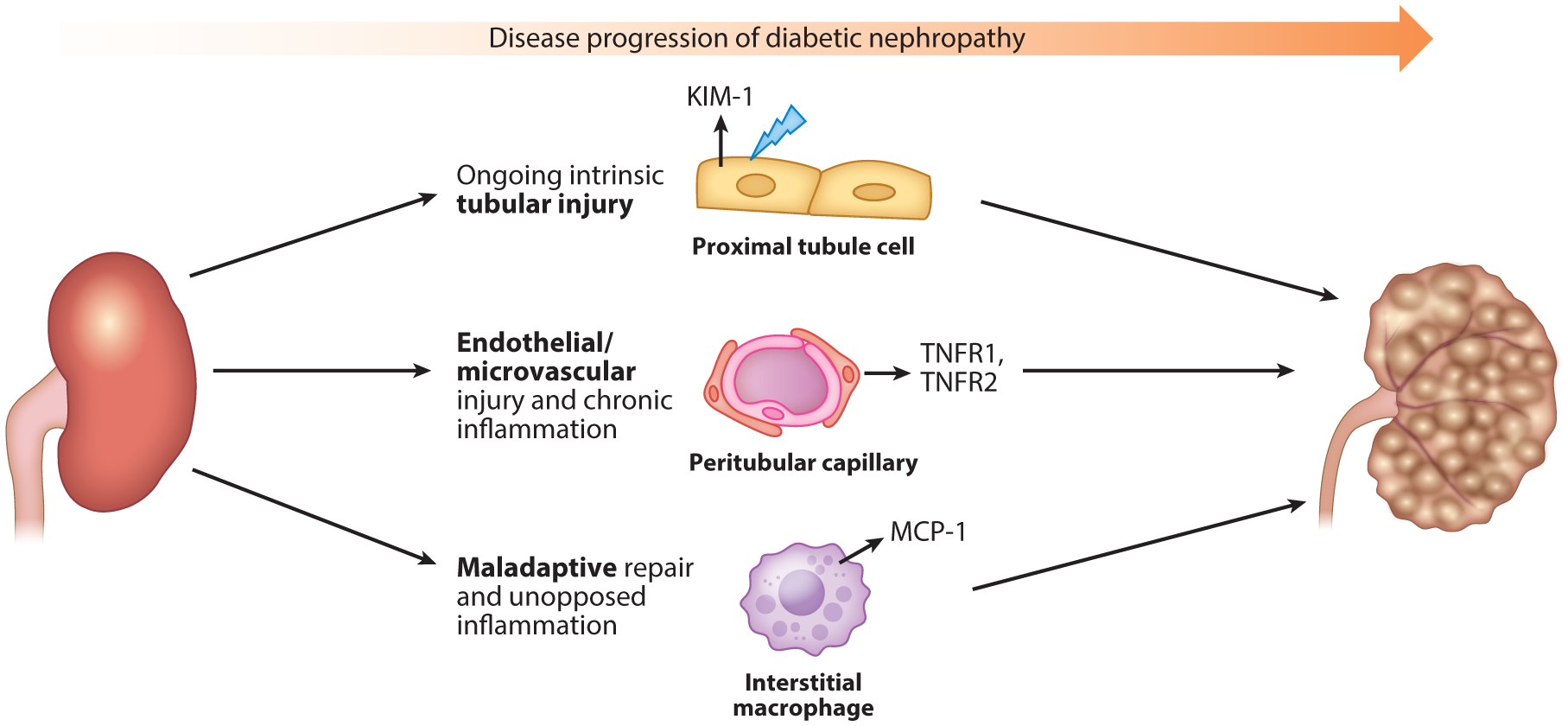
The current unidimensional paradigm of kidney disease detection is incompatible with the complexity and heterogeneity of renal pathology. The diagnosis of kidney disease has largely focused on glomerular filtration, while assessment of kidney tubular health has notably been absent. Following insult, the kidney tubular cells undergo a cascade of cellular responses that result in the production and accumulation of low-molecular-weight proteins in the urine and systemic circulation. Modern advancements in molecular analysis and proteomics have allowed the identification and quantification of these proteins as biomarkers for assessing and characterizing kidney diseases. In this review, we highlight promising biomarkers of kidney tubular health that have strong underpinnings in the pathophysiology of kidney disease. These biomarkers have been applied to various specific clinical settings from the spectrum of acute to chronic kidney diseases, demonstrating the potential to improve patient care.

Keywords: AKI; CKD; acute kidney injury; biomarkers; chronic kidney disease; kidney.


Figures

Figure 1
Figure 1
Biomarkers of kidney tubular health by anatomic localization and pathophysiology. Biomarkers of kidney tubular health are labeled corresponding to the anatomic site and/or mechanism of production. Abbreviations: EGF, epidermal growth factor; IGFBP-7, insulin-like growth factor-binding protein 7; IL, interleukin; KIM-1, kidney injury molecule-1; L-FABP, liver-type fatty acid–binding-protein; MCP-1, monocyte chemoattractant protein-1; NGAL, neutrophil gelatinase-associated lipocalin; PIIINP, procollagen type III N-terminal propeptide; TIMP-2, tissue inhibitor of metalloproteinases-2; UMOD, uromodulin; YKL-40, chitinase 3-like protein 1. Figure adapted with permission from Reference . Copyright 2013, American Society of Nephrology.
Figure 2
Figure 2
Biomarkers of AKI. The upper panel contains an AKI biopsy specimen, and the bottom panel is a close-up graphical representation of the portion of the AKI biopsy that is boxed in black and depicts three domains of kidney biomarkers. Biomarkers of tubular injury that are depicted include KIM-1 and NGAL, which are produced by proximal tubular cells early after kidney injury. Biomarkers of tubular function that are depicted include L-FABP, CysC, and α1M, which are proteins that build up in the urine due to tubular dysfunction. Biomarkers of inflammation that are depicted include IL-6, IL-10, IL-18, and MCP-1, which are produced as part of the inflammatory response to injury. Abbreviations: AKI, acute kidney injury; α1M, alpha-1 microglobulin; CysC, cystatin C; IL, interleukin; KIM-1, kidney injury molecule-1; L-FABP, liver-type fatty acid-binding-protein; MCP-1, monocyte chemoattractant protein-1; NGAL, neutrophil gelatinase-associated lipocalin.
Figure 3
Figure 3
Distinguishing elevations of serum creatinine with biomarkers of kidney tubular health. Elevations in serum creatinine that meet the clinical definition of AKI do not necessarily represent true injury and have differing significance and clinical implications depending on the clinical milieu. Biomarker changes, their significance in elucidating the meaning of seemingly uniform elevations of serum creatinine, and clinical implications for patient management are detailed for the settings of postcardiac surgery, cardiorenal syndrome, hepatorenal syndrome, and kidney transplantation. Abbreviations: AKI, acute kidney injury; IL, interleukin; KIM-1, kidney injury molecule-1; NGAL, neutrophil gelatinase-associated lipocalin.
Figure 4
Figure 4
Opportunities for biomarkers of kidney tubular health to advance care in CKD. The left-hand y-axis depicts the eGFR, and the red line represents the eGFR trajectory with respect to time over the disease course. The right-hand y-axis depicts the number of remaining nephrons in millions, and the purple line depicts the nephron number with respect to time over the disease course. The delay in diagnosis (labeled as the distance between the dashed green vertical lines) may be reduced by use of biomarkers of kidney tubular health, which begin to increase as the nephron number begins to fall. In the earliest stages of CKD, the number of nephrons begins to decrease prior to being captured by clinically meaningful decreases in eGFR, as the kidney is able to compensate for loss of nephrons via the phenomenon of renal filtration reserve. Trapezoid A is labeled as renal reserve and encompasses the early course of disease when nephron numbers have begun to fall, yet eGFR is stable or only beginning to decline but is still above the threshold for clinical classification of CKD (dashed black horizontal line at eGFR of 90 mL/min/1.73 m2). In this range, novel biomarkers may address the critical limitations of serum creatinine and eGFR by detecting the earliest stages of kidney damage of incident CKD. Trapezoid B encompasses a region in which the effects of renal reserve may persist but are tapering out. In this range, decreases in nephron number may be paralleled by decreases in eGFR; thus, while biomarkers may be increasing and signaling intrinsic kidney damage, the kidney may also manifest loss of nephron number as commensurate eGFR declines because of the loss of renal reserve at this point. Thus, biomarkers may have less utility at this stage in the progression of CKD in discerning injury to the kidney; however, they may be able to address other limitations of serum creatinine. Triangle C represents advanced stages of CKD, in which the current therapeutic window for intervention lies. Utilizing biomarkers of tubular injury may allow for identification of therapeutic targets and shift this window to earlier points in the disease course. Abbreviations: CKD, chronic kidney disease; eGFR, estimated glomerular filtration rate.
Figure 5
Figure 5
Distinct mechanisms of diabetic nephropathy progression. Diabetic nephropathy is a heterogeneous condition that may involve diverse mechanisms. This may explain why a portion of patients with diabetic kidney disease continue to experience disease progression, despite optimal therapy with the current standard of treatments, including angiotensin-converting enzyme inhibitors and angiotensin II receptor blockers. These current therapies do not address the underlying mechanisms that drive the progression of diabetic kidney disease, which may include ongoing intrinsic tubular injury, manifested by elevated levels of KIM-1; endothelial/microvascular injury and chronic inflammation, manifested by elevated levels of TNFR1 and TNFR2; and maladaptive repair and unopposed inflammation, manifested by elevated levels of MCP-1. These biomarkers may discern distinct pathways of injury and inform therapeutic interventions for diabetic nephropathy. Abbreviations: KIM-1, kidney injury molecule-1; MCP-1, monocyte chemoattractant protein-1; TNFR, tumor necrosis factor receptor.
Figure 6
Figure 6
Application of kidney biomarkers in clinical trials. Biomarkers of tubular health may be applied in various stages of clinical trials. At enrollment, diagnostic biomarkers (red) can be used to identify participants with true intrinsic tubular injury; prognostic biomarkers (orange) can be used to identify participants at high risk for outcomes; and predictive biomarkers (green) can be used to identify participants likely to respond to an intervention of interest. By using these biomarkers to assess eligibility and exclude individuals who may not be truly suitable for the study despite an elevation in serum creatinine, investigators may be able to conduct more efficient, cost-effective trials. In the follow-up phase of trials, pharmacodynamics biomarkers (purple) may be used to assess safety of interventions and serve as surrogate outcomes for kidney damage. With the wide breath of biomarkers now available, biomarkers may be combined with novel statistical techniques in the analysis phase. Figure adapted with permission from Reference .

References

    1. Uchino S, Kellum JA, Bellomo R, Doig GS, Morimatsu H, et al. 2005. Acute renal failure in critically ill patients: a multinational, multicenter study. JAMA 294:813–18 - PubMed
    1. Kampmann J, Siersbaek-Nielsen K, Kristensen M, Hansen JM. 1974. Rapid evaluation of creatinine clearance. Acta Med. Scand 196:517–20 - PubMed
    1. Nickolas TL, Barasch J, Devarajan P. 2008. Biomarkers in acute and chronic kidney disease. Curr. Opin. Nephrol. Hypertens 17:127–32 - PubMed
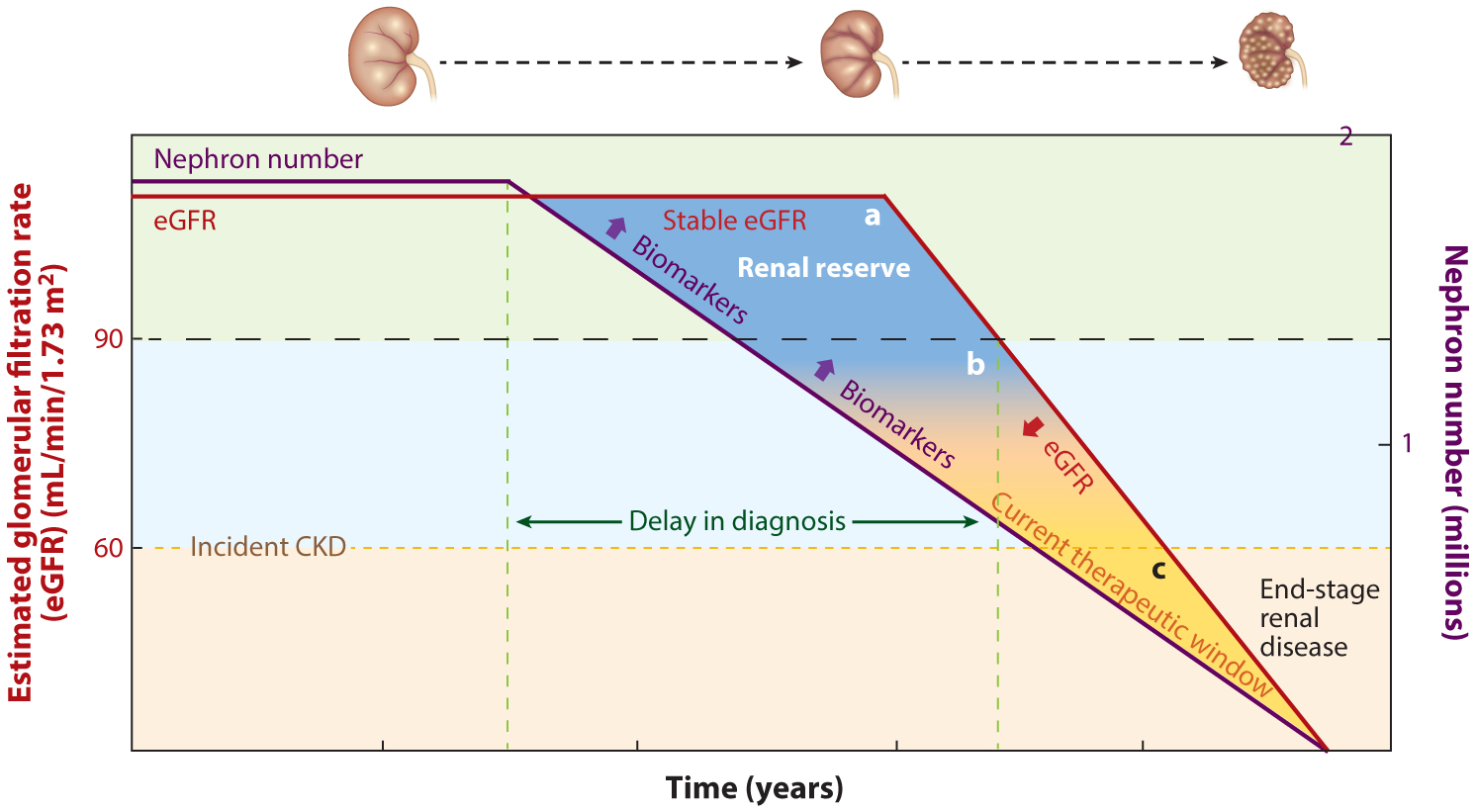
    1. Bosch JP. 1995. Renal reserve: a functional view of glomerular filtration rate. Semin. Nephrol 15:381–85 - PubMed
    1. Herrera J, Rodríguez-Iturbe B. 1998. Stimulation of tubular secretion of creatinine in health and in conditions associated with reduced nephron mass. Evidence for a tubular functional reserve. Nephrol. Dial. Transplant 13:623–29 - PubMed

Publication types