Review
. 2020 Mar;1867(3):118436.
doi: 10.1016/j.bbamcr.2019.01.017. Epub 2019 Feb 8.

Mix and (mis-)match - The mechanosensing machinery in the changing environment of the developing, healthy adult and diseased heart

Affiliations
Review

Mix and (mis-)match - The mechanosensing machinery in the changing environment of the developing, healthy adult and diseased heart

Matthew Ward et al. Biochim Biophys Acta Mol Cell Res. 2020 Mar.

Abstract

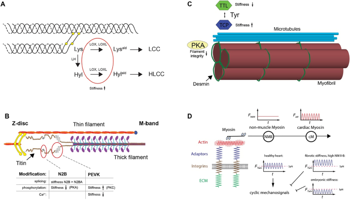
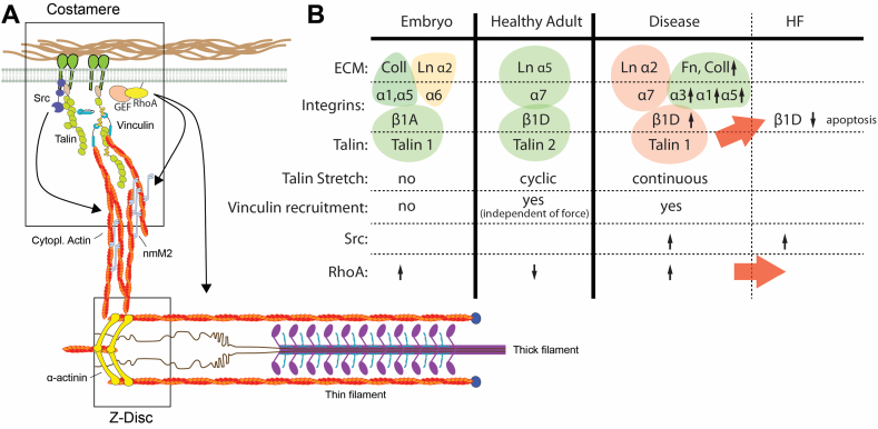
The composition and the stiffness of cardiac microenvironment change during development and/or in heart disease. Cardiomyocytes (CMs) and their progenitors sense these changes, which decides over the cell fate and can trigger CM (progenitor) proliferation, differentiation, de-differentiation or death. The field of mechanobiology has seen a constant increase in output that also includes a wealth of new studies specific to cardiac or cardiomyocyte mechanosensing. As a result, mechanosensing and transduction in the heart is increasingly being recognised as a main driver of regulating the heart formation and function. Recent work has for instance focused on measuring the molecular, physical and mechanical changes of the cellular environment - as well as intracellular contributors to the passive stiffness of the heart. On the other hand, a variety of new studies shed light into the molecular machinery that allow the cardiomyocytes to sense these properties. Here we want to discuss the recent work on this topic, but also specifically focus on how the different components are regulated at various stages during development, in health or disease in order to highlight changes that might contribute to disease progression and heart failure.


Figures

Fig. 1
Fig. 1
Main contributors to the passive stiffness in the heart. A) Collagen is crosslinked through lysyl oxidases (LOX, LOXL), increasing its stiffness. Additionally, the stability of the crosslinks is determined through activity of lysyl hydroxylases (LH). B) The stiffness of the elastic N2B and the PEVK domains of titin can be changed through splicing, phosphorylation or calcium binding. C) Detyrosination (through unknown tubulin carboxypeptidases) of microtubules leads to crosslinking with desmin and high-resistance buckling as opposed to low resistance sliding behavior. Detyrosination can be reversed by tubulin tyrosine ligase (TTL) activity. D) Non-muscle myosin can increase the basal tension that is sensed by cardiomyocyte integrin adhesions, thereby changing the mechanical signalling through adaptor proteins, such as talin (e.g. cyclic vs continuous stretching of talin) – figure adapted from Pandey et al., 2018 [7].
Fig. 2
Fig. 2
Changing components of the mechanosensing apparatus. A) Schematic of the integrin adhesions in the costameres and their connection to the sarcomere. Note only a few relevant components are displayed and proteins are not in scale. B) Overview of the different isoforms for ECM proteins, integrins and talin, as well as different downstream effects in embryonic, healthy adult and diseased hearts. Red arrows indicate events that can lead to progression to heart failure, i.e. the downregulation of β1D integrin through talin 1 overexpression, leading to apoptosis and heart failure; and heightened RhoA activity, which is also associated with heart failure. (For interpretation of the references to colour in this figure legend, the reader is referred to the web version of this article.)

References

    1. Iskratsch T., Wolfenson H., Sheetz M.P. Appreciating force and shape-the rise of mechanotransduction in cell biology. Nat. Rev. Mol. Cell Biol. 2014;15:825–833. - PMC - PubMed
    1. Emery J.L., Mithal A. Weights of cardiac ventricles at and after birth. Br. Heart J. 1961;23:313. - PMC - PubMed
    1. Romero T., Covell J., Friedman W.F. A comparison of pressure-volume relations of the fetal, newborn, and adult heart. Am. J. Physiol. 1972;222:1285–1290. - PubMed
    1. Rudolph A.M. The changes in the circulation after birth. Circulation. 1970;41:343–359. - PubMed
    1. Andres-Delgado L., Mercader N. Interplay between cardiac function and heart development. Biochim. Biophys. Acta. 2016;1863:1707–1716. - PMC - PubMed

Publication types

MeSH terms