. 2019 Apr:134:604-616.
doi: 10.1016/j.freeradbiomed.2019.02.001. Epub 2019 Feb 10.

The molecular chaperone sigma 1 receptor mediates rescue of retinal cone photoreceptor cells via modulation of NRF2

Affiliations

The molecular chaperone sigma 1 receptor mediates rescue of retinal cone photoreceptor cells via modulation of NRF2

J Wang et al. Free Radic Biol Med. 2019 Apr.

Abstract

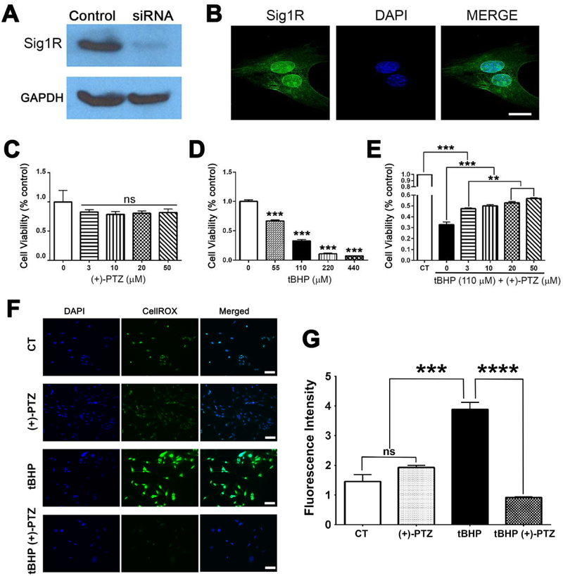
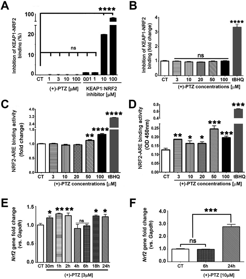
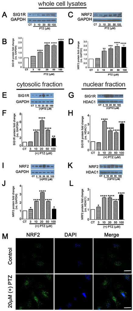
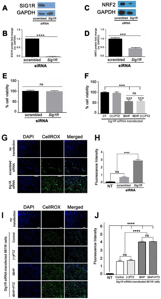
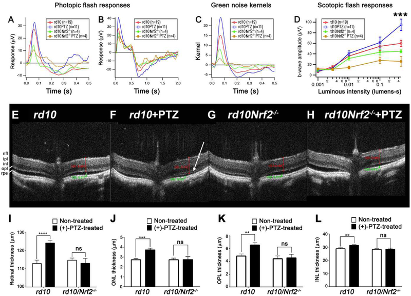
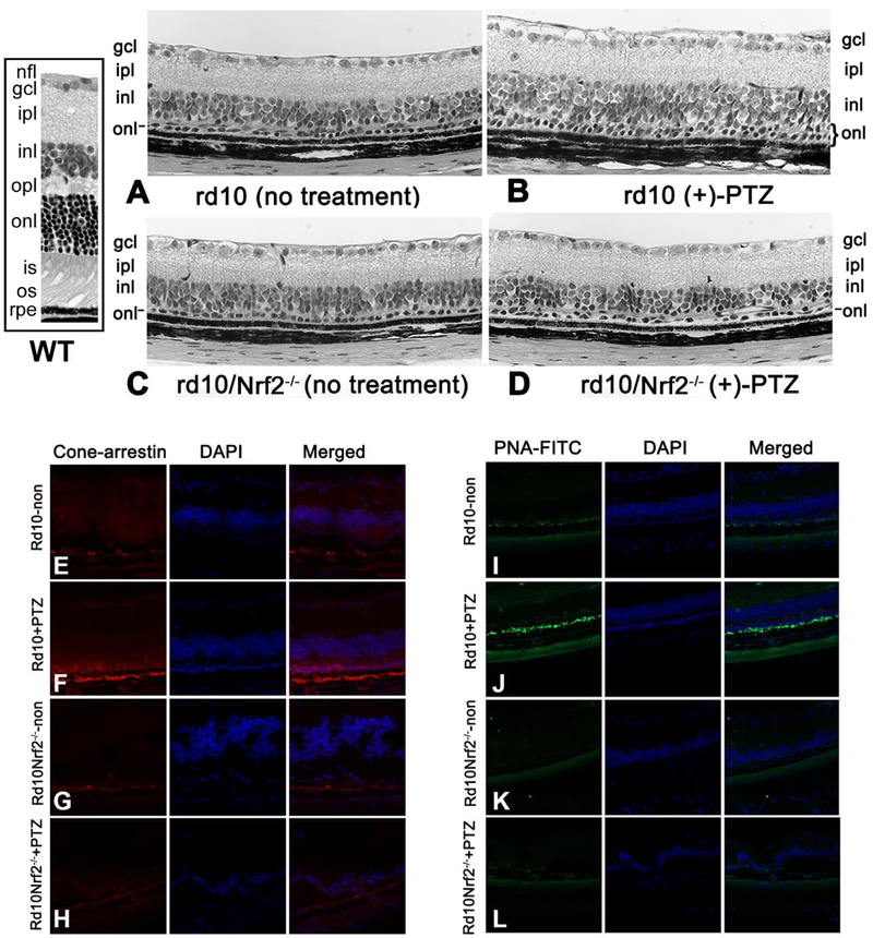
Sigma 1 receptor (Sig1R), a putative molecular chaperone, has emerged as a novel therapeutic target for retinal degenerative disease. Earlier studies showed that activation of Sig1R via the high-affinity ligand (+)-pentazocine ((+)-PTZ) induced profound rescue of cone photoreceptor cells in the rd10 mouse model of retinitis pigmentosa; however the mechanism of rescue is unknown. Improved cone function in (+)-PTZ-treated mice was accompanied by reduced oxidative stress and normalization of levels of NRF2, a transcription factor that activates antioxidant response elements (AREs) of hundreds of cytoprotective genes. Here, we tested the hypothesis that modulation of NRF2 is central to Sig1R-mediated cone rescue. Activation of Sig1R in 661W cone cells using (+)-PTZ induced dose-dependent increases in NRF2-ARE binding activity and NRF2 gene/protein expression, whereas silencing Sig1R significantly decreased NRF2 protein levels and increased oxidative stress, although (+)-PTZ did not disrupt NRF2-KEAP1 binding. In vivo studies were conducted to investigate whether, in the absence of NRF2, activation of Sig1R rescues cones. (+)-PTZ was administered systemically for several weeks to rd10/nrf2+/+ and rd10/nrf2-/- mice. Through post-natal day 42, cone function was significant in rd10/nrf2+/+, but minimal in rd10/nrf2-/- mice as indicated by electroretinographic recordings using natural noise stimuli, optical coherence tomography and retinal histological analyses. Immunodetection of cones was limited in (+)-PTZ-treated rd10/nrf2-/-, though considerable in (+)-PTZ-treated rd10/nrf2+/+mice. The data suggest that Sig1R-mediated cone rescue requires NRF2 and provide evidence for a previously-unrecognized relationship between these proteins.

Keywords: NRF2-KEAP1; NRF2-Neh luciferase assay; Oxidative stress; Retina; Retinal neuroprotection; Retinitis pigmentosa; rd10 mouse.


Figures

Fig. 1.
Fig. 1.. Activation of Sig1R enhances viability and attenuates oxidative stress.
(A) Immunoblotting to detect SIG1R was performed in 661W cells using a rabbit polyclonal antibody. A band of the expected size (27kD Mr) was detected in 661W cells (control); there was only a faint band detected in 661W cells in which Sig1R gene expression had been silenced using siRNA technology; GAPDH was used as the protein loading control. (B) Immunofluorescence in 661W cells detected Sig1R (green fluorescence), especially in the nucleus and nuclear membrane, the nucleus is labeled with DAPI (blue fluorescence). The viability of 661W cells incubated 24 h with (C) increasing concentrations of (+)-PTZ [0-50μM]; (D) increasing concentrations of tBHP [0-440μM]; or (E) 110μM tBHP in the presence of increasing concentrations of (+)-PTZ [0-50μM] was assessed using the MTT assay. (F) 661W cells were seeded on coverslips for 18 h. Cells either were or were not (control) exposed for 3 h to 110μM tBHP in the presence or absence of (+)-PTZ, including cells treated with (+)-PTZ alone. They were incubated with CellROX® Green Reagent to detect ROS; green fluorescent signals indicating ROS were visualized by epifluorescence; DAPI was used to label nuclei (blue). Scale bar, 50 μm. (G) Quantification of fluorescent intensity reflecting ROS levels of data shown in panel F. Data are presented as mean ± standard error of the mean (SEM). Data represent three independent experiments performed in duplicate. Significant differences are indicated: ** p<0.01, *** p<0.001****p<0.0001. (CT = control, (+)-PTZ = (+)-pentazocine, tBHP = tertiary butyl hydroperoxide).
Fig. 2.
Fig. 2.. Activation of Sig1R does not disrupt KEAP1-NRF2 binding directly, but does increase NRF2-ARE binding and Nrf2 expression.
661W cells were exposed to varying concentrations of (+)-PTZ after which KEAP1-NRF2 binding was analyzed using two methods: (A) fluorescence polarization-based detection assay, and (B) Neh2-Luciferase reporter assay. 661W cells were exposed to varying concentrations of (+)-PTZ and NRF2-ARE binding activity was analyzed using two methods: (C) AREc32 cells stably expressing ARE-Luc and (D) nuclear extracts from 661W cells using TransAM NRF2 kit. 661W cells were exposed (E) to (+)-PTZ [3μM] over a 24 h time course; and (F) to (+)-PTZ [10μM] for 6 and 24 h following which the expression of Nrf2 was determined by qRT-PCR using primer pairs specific for Nrf2. Data are mean ± SEM of triplicate or quadruplicate measurements *p< 0.05. ** p< 0.01; *** p< 0.001, **** p< 0.0001. (CT = control, (+)-PTZ = (+)-pentazocine, tBHQ = tertiary butyl hydroquinone).
Fig. 3.
Fig. 3.. Sig1R activation increases NRF2 protein level.
661W cells were exposed 24 h to varying concentrations of (+)-PTZ; levels of SIG1R and NRF2 proteins in whole cell lysates (A-D) or in cytosolic/nuclear fractions (E-L) were determined by western blotting. Band densities were quantified using ImageJ and expressed as fold change compared to GAPDH. Representative western blots for (A) SIG1R (C) NRF2 in whole cell lysates and graphical depiction of fold change (protein versus GAPDH) for (B) SIG1R, (D) NRF2. Cytosolic and nuclear protein fractions were prepared and extracts were subjected to western blot analysis to evaluate levels of (E) cytosolic SIG1R, (G) nuclear SIG1R, (I) cytosolic NRF2; (K) nuclear NRF2. Each experiment was performed in triplicate. Band density was quantified as a ratio to GAPDH for the cytosolic fractions of (F) SIG1R and (H) NRF2, and as a ratio to HDAC1 for the nuclear fractions of (J) SIG1R and (L) NRF2. Data are mean ± SEM of triplicate measurements, *p< 0.05. ** p< 0.01; *** p< 0.001, **** p< 0.0001, ns = not significant. (CT = control, (+)-PTZ = (+)-pentazocine). (M) Immunofluorescent detection of NRF2 (green fluorescence) in 661W cells treated with 20μM (+)-PTZ, the nucleus is labeled with DAPI (blue fluorescence). Calibration bar = 20μm.
Fig. 4.
Fig. 4.. NRF2 levels are attenuated and oxidative stress is increased in 661W cells in which Sig1R has been silenced.
(A) Immunoblotting to detect SIG1R in 661W cells transfected with either “scrambled” siRNA or Sig1R siRNA. (B) Quantitation of multiple repetitions of the experiment shown in A. (C) Immunoblotting to detect NRF2 in 661W cells in cells transfected with either “scrambled” siRNA or Sig1R siRNA. (D) Quantitation of multiple repetitions of the experiment shown in C. (E) Cell viability was assessed using the MTT assay in 661W cells 3 days after transfection with either “scrambled” siRNA or Sig1R siRNA; there was no significant difference in viability between the two groups. 3 days after transfection with Sig1R siRNA, 661W cells were treated 18h with 110μM tBHP in the presence/absence of PTZ ([10μM], 30 min pretreatment followed by co-treatment). Cell viability was assessed using the MTT assay. Data were analyzed using one-way ANOVA and in cases of multiple comparisons (F) the Tukey-Kramer post-hoc test for multiple comparisons. (G) Cells were incubated with CellROX® Green Reagent to detect ROS; green fluorescent signals indicating ROS were visualized by epifluorescence; DAPI was used to label nuclei (blue). Scale bar, 100 μm. (H) Quantification of fluorescent intensity reflecting ROS levels of data shown in panel G. (I) Cells transfected as described for panel A were exposed to 110 μM tBHP, a known oxidative stress inducer in the presence or (+)-PTZ [10μM] and oxidative stress was assessed using CellROX® Green Reagent. (J) Quantification of fluorescent intensity reflecting ROS levels of data shown in panel (B). Data are mean ± SEM of triplicate measurements *** p< 0.001; ****p<0.0001. (CT = control, (+)-PTZ = (+)-pentazocine, tBHP = tertiary butyl hydroperoxide, ns = not significant).
Fig. 5.
Fig. 5.. Photopic ERG responses and in vivo retinal structure are not improved in rd10/Nrf2−/− mice treated with (+)-PTZ.
Photopic ERG responses were performed on four groups of mice: (1) rdl0 mice receiving no treatment, (2) rdl0 mice administered (+)-PTZ (rdl0+PTZ), (3) rdl0/Nrf2−/− mice receiving no treatment, (4) rd10/Nrf2−/− mice administered (+)-PTZ (rd10/Nrf2−/−+PTZ). Averaged responses to photopic flashes are shown for (A) 0.5 sec and (B) 2.0 sec testing periods. (C) Averaged kernels derived from responses to natural noise stimuli. (D) Averaged scotopic ERG responses to 5 ms flashes at a series of intensities for rd10, rd10+PTZ, rd10/Nrf2−/−, rd10/Nrf2−/−+PTZ. Intensities are in units of candela-seconds per meter squared. (***p=0.002 for rd10+PTZ at highest intensity compared to all other groups). Panels E-H: Representative SD-OCT data obtained from rd10, rd10+PTZ, rd10/Nrf2−/−, rd10/Nrf2−/−+PTZ at P42. (I-L) Data from segmentation analysis for (I) total retinal thickness; (J) outer nuclear layer (ONL) thickness; (K) outer plexiform layer (OPL) thickness, (L) inner nuclear layer (INL) thickness. OCT data are the mean ± SEM of analyses in 7-12 mice per group (Table S1). Significance: *p < 0.05,**p < 0.01, ***p < 0.001 ****p < 0.0001. (nfl = nerve fiber layer, ipl = inner plexiform layer, inl = inner nuclear layer, opl = outer plexiform layer, rpe = retinal pigment epithelium). Note: The onl (outer nuclear layer) is visible in the rd10+PTZ mouse retina (F) as indicated by white arrow, but is not visible in retinas of mice in the other 3 groups.
Fig. 6.
Fig. 6.. Cone photoreceptor cells are not rescued in rd10/Nrf2−/− mice treated with (+)-PTZ.
Retinal sections of eyes embedded in JB-4 and stained with H&E from a WT mouse (boxed side panel) and four groups of mice: (A) rd10 mice receiving no treatment, (B) rd10 mice administered (+)-PTZ (rd10+PTZ), (C) rd10/Nrf2−/− mice receiving no treatment, (D) rd10/Nrf2−/− mice administered (+)-PTZ (rd10/Nrf2−/−+PTZ). Note that two rows of photoreceptor cells are present in the rd10+PTZ retinas, but not in the other mouse groups. The sections are representative of the retinas analyzed; the numbers of mice evaluated are provided in Suppl. Table 1. Additional eyes were harvested from the four groups and processed for cryosectioning to detect cone-arrestin or PNA-FITC, both markers of cone photoreceptors. Cone arrestin labeling for (E) rd10-no treatment, (F) rd10+PTZ, (G) rd10/nrf2−/−-no treatment, (H) rd10/nrf2−/−+PTZ; PNA-FITC labeling for (I) rd10-no treatment, (J) rd10+PTZ, (K) rd10/nrf2−/−-no treatment, (L) rd10/nrf2−/−+PTZ. Abbreviations: nfl, nerve fiber layer; gcl, ganglion cell layer, ipl, inner plexiform layer; inl, inner nuclear layer; opl, outer plexiform layer; onl, outer nuclear layer; is, inner segment; os, outer segment; rpe, retinal pigment epithelium.

Similar articles

Cited by

References

    1. Schmidt HR, Zheng S, Gurpinar E, Koehl A, Manglik A, Kruse AC. Crystal structure of the human σ1 receptor. Nature. 532:527–30;2016. - PMC - PubMed
    1. Su TP, Su TC, Nakamura Y, Tsai SY. The Sigma-1 Receptor as a Pluripotent Modulator in Living Systems. Trends Pharmacol Sci. 37:262–278;2016. - PMC - PubMed
    1. Tadić V, Malci A, Goldhammer N, Stubendorff B, Sengupta S, Prell T, Keiner S, Liu J, Guenther M, Frahm C, Witte OW, Grosskreutz J. Sigma 1 receptor activation modifies intracellular calcium exchange in the G93A(hSOD1) ALS model. Neuroscience. 359:105–118;2017. - PubMed
    1. Mavlyutov TA, Baker EM, Losenegger TM, Kim JR, Torres B, Epstein ML, Ruoho AE. The Sigma-1 Receptor-A Therapeutic Target for the Treatment of ALS? Adv Exp Med Biol. 964:255–265;2017. - PubMed
    1. Mancuso R, Oliván S, Rando A, Casas C, Osta R, Navarro X. Sigma-1R agonist improves motor function and motoneuron survival in ALS mice. Neurotherapeutics. 9:814–26;2012. - PMC - PubMed

Publication types

MeSH terms

LinkOut - more resources