. 2019 Feb 8;20(3):721.
doi: 10.3390/ijms20030721.

The Composition of Hyperacute Serum and Platelet-Rich Plasma Is Markedly Different despite the Similar Production Method

Affiliations

The Composition of Hyperacute Serum and Platelet-Rich Plasma Is Markedly Different despite the Similar Production Method

Dorottya Kardos et al. Int J Mol Sci. .

Abstract

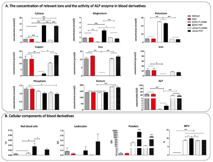
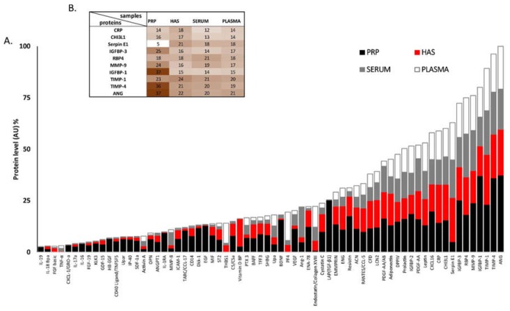
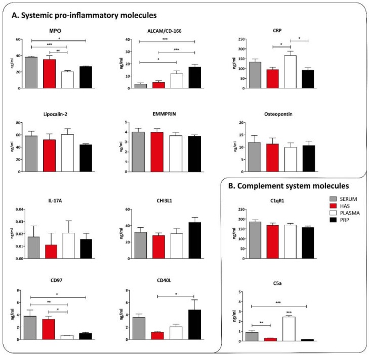
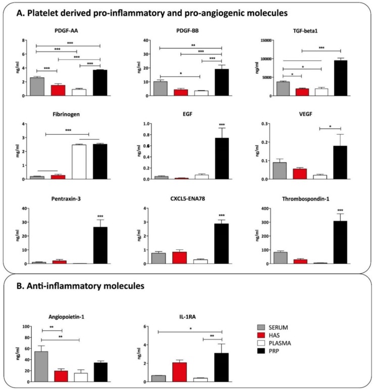
Autologous blood derived products, such as platelet-rich plasma (PRP) and platelet-rich fibrin (PRF) are widely applied in regenerative therapies, in contrast to the drawbacks in their application, mainly deriving from the preparation methods used. Eliminating the disadvantages of both PRP and PRF, hyperacute serum (HAS) opens a new path in autologous serum therapy showing similar or even improved regenerative potential at the same time. Despite the frequent experimental and clinical use of PRP and HAS, their protein composition has not been examined thoroughly yet. Thus, we investigated and compared the composition of HAS, serum, PRP and plasma products using citrate and EDTA by simple laboratory tests, and we compared the composition of HAS, serum, EDTA PRP and plasma by Proteome Profiler and ELISA assays. According to our results the natural ionic balance was upset in both EDTA and citrate PRP as well as in plasma. EDTA PRP contained significantly higher level of growth factors and cytokines, especially platelet derived angiogenic and inflammatory proteins, that can be explained by the significantly higher number of platelets in EDTA PRP. The composition analysis of blood derivatives revealed that although the preparation method of PRP and HAS were similar, the ionic and protein composition of HAS could be advantageous for cell function.

Keywords: blood derived products; composition; hyperacute serum; platelet-rich plasma.


Conflict of interest statement

Z.L. owns stock in OrthoSera GmbH, a startup company developing the hyperacute serum technology towards clinical application.

Figures

Figure 1
Figure 1
Quantitative determination of the concentration of relevant ions and the activity of alkaline phosphatase (ALP) enzyme (A) and the number of red blood cells, leukocytes and platelets in the serum and plasma fractions (B), n = 4. The significance level was p > 0.05, where * means that p is between 0.01 and 0.05, ** means that p is between 0.01 and 0.001, and *** means that p is lower than 0.001.
Figure 2
Figure 2
Semi-quantitative Proteome Profiler analysis of serum, hyperacute serum (HAS), plasma, and platelet-rich fibrin (PRP). On the bar chart proteins exceeding 2% (AU) of the total protein content (A) are presented. The level of the top 10 angiogenic proteins and cytokines are presented on a heat map. (B) The level of the proteins is expressed in % compared to the combined arbitrary unit of ANG that was considered to be 100%. n = 8.
Figure 3
Figure 3
Concentration of systemic pro-inflammatory molecules (A) and complement system related molecules (B) in blood derivatives, n = 8. The significance level was p > 0.05, where * means that p is between 0.01 and 0.05, ** means that p is between 0.01 and 0.001, and *** means that p is lower than 0.001.
Figure 4
Figure 4
The concentration of platelet-derived inflammatory molecules (A) and anti-inflammatory molecules (B) in blood derivatives, n = 8. The significance level was p > 0.05, where * means that p is between 0.01 and 0.05, ** means that p is between 0.01 and 0.001, and *** means that p is lower than 0.001.
Figure 5
Figure 5
For serum isolation, whole blood was obtained from donors in VACUETTE® 9 mL Z Serum C/A tubes (Greiner Bio-One, Kremsmünster, Austria). Blood was allowed to clot for 30 min (a) and centrifuged at 1710× g for 5 min at room temperature (b). The supernatant formed after centrifugation is called serum (c,d).
Figure 6
Figure 6
For hyperacute serum isolation, a whole blood sample was obtained from healthy donors (28–45 years) in VACUETTE® 9 mL Z Serum C/A tubes (Greiner Bio-One) (a) and it was immediately centrifuged at 1710× g for 5 min at room temperature (b). After centrifugation two layers were formed in the tubes. The top layer was the platelet-rich fibrin clot and the bottom layer contains red blood cells (c). PRF (platelet-rich fibrin) as removed using sterile forceps in a biosafety cabinet (d), red blood cells at the bottom of the fibrin clot were cut away (e) and the clot was placed onto a 110 mm long, 75 mm wide custom-made plastic grid with 5 mm diameter holes on it. It was sterilized in an autoclave before use (f). The hyperacute serum was squeezed out from the PRF clot using a sterile spatula (g,h) [24].
Figure 7
Figure 7
For PRP isolation, whole blood was obtained from donors in VACUETTE® 9 mL K3 EDTA blood collection tubes (Greiner Bio-One) and VACUTTE 3.5 mL sodium citrate 3.2% blood collection tubes (Greiner Bio-One) (a) then centrifuged at 1710× g for 5 min at room temperature (b). The supernatant formed after centrifugation is called plasma (c,d).
Figure 8
Figure 8
For PRP isolation, whole blood was obtained from donors in VACUETTE® 9 mL K3 EDTA blood collection tubes (Greiner Bio-One) and VACUTTE 3.5 mL sodium citrate 3.2% blood collection tubes (Greiner Bio-One) (a) then centrifuged at 320 g for 12 min at room temperature (b). The platelet-rich layer above the buffy coat was aspirated and transferred into a 15 mL tube (c,d) and centrifuged at 1710× g for 10 minutes (e). The resulting platelet pellet was resuspended in the same volume as the isolated hyperacute serum from the same donor (f). PRP was activated by 10 IU thrombin and 10 mg calcium-chloride (Sigma-Aldrich, St. Louis, MO, USA) in case of both EDTA and citrate tubes (g,h).

References

    1. Fotouhi A., Maleki A., Dolati S., Aghebati-Maleki A., Aghebati-Maleki L. Platelet rich plasma, stromal vascular fraction and autologous conditioned serum in treatment of knee osteoarthritis. Biomed. Pharmacother. 2018;104:652–660. doi: 10.1016/j.biopha.2018.05.019. - DOI - PubMed
    1. Le A.D.K., Enweze L., DeBaun M.R., Dragoo J.L. Current Clinical Recommendations for Use of Platelet-Rich Plasma. Curr. Rew. Musculoskelet. Med. 2018;11:624–634. doi: 10.1007/s12178-018-9527-7. - DOI - PMC - PubMed
    1. Amable P.R., Carias R.B.V., Teixeira M.V.T., da Cruz Pacheco I., Corrêa do Amaral R.J.F., Granjeiro J.M., Borojevic R. Platelet-rich plasma preparation for regenerative medicine: Optimization and quantification of cytokines and growth factors. Stem Cell Res. Ther. 2013;4:67. doi: 10.1186/scrt218. - DOI - PMC - PubMed
    1. Prakash S., Thakur A. Platelet concentrates: Past, present and future. J. Oral Maxillofac. Surg. 2011;10:45–49. doi: 10.1007/s12663-011-0182-4. - DOI - PMC - PubMed
    1. Andia I., Maffulli N. Use of platelet-rich plasma for patellar tendon and medial collateral ligament injuries: Best current clinical practice. J. KNEE Surg. 2015;28:11–18. doi: 10.1055/s-0034-1384671. - DOI - PubMed