An Approach to Automatic Hard Exudate Detection in Retina Color Images by a Telemedicine System Based on the d-Eye Sensor and Image Processing Algorithms
- PMID: 30744032
- PMCID: PMC6387053
- DOI: 10.3390/s19030695
An Approach to Automatic Hard Exudate Detection in Retina Color Images by a Telemedicine System Based on the d-Eye Sensor and Image Processing Algorithms
Abstract
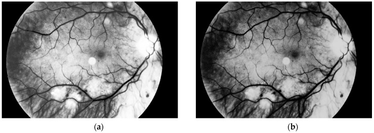
Hard exudates are one of the most characteristic and dangerous signs of diabetic retinopathy. They can be marked during the routine ophthalmological examination and seen in color fundus photographs (i.e., using a fundus camera). The purpose of this paper is to introduce an algorithm that can extract pathological changes (i.e., hard exudates) in diabetic retinopathy. This was a retrospective, nonrandomized study. A total of 100 photos were included in the analysis-50 sick and 50 normal eyes. Small lesions in diabetic retinopathy could be automatically diagnosed by the system with an accuracy of 98%. During the experiments, the authors used classical image processing methods such as binarization or median filtration, and data was read from the d-Eye sensor. Sixty-seven patients (39 females and 28 males with ages ranging between 50 and 64) were examined. The results have shown that the proposed solution accuracy level equals 98%. Moreover, the algorithm returns correct classification decisions for high quality images and low quality samples. Furthermore, we consider taking retina photos using mobile phones rather than fundus cameras, which is more practical. The paper presents an innovative approach. The results are introduced and the algorithm is described.
Keywords: automatic diagnosis; d-eye sensor; hard exudates; image processing; retina.
Conflict of interest statement
The authors declare no conflicts of interest.
Figures


Similar articles
-
Detection of Hard Exudates Using Evolutionary Feature Selection in Retinal Fundus Images.J Med Syst. 2019 May 29;43(7):209. doi: 10.1007/s10916-019-1349-7. J Med Syst. 2019. PMID: 31144041
-
A novel automatic image processing algorithm for detection of hard exudates based on retinal image analysis.Med Eng Phys. 2008 Apr;30(3):350-7. doi: 10.1016/j.medengphy.2007.04.010. Epub 2007 Jun 6. Med Eng Phys. 2008. PMID: 17556004
-
Decision support system for the detection and grading of hard exudates from color fundus photographs.J Biomed Opt. 2011 Nov;16(11):116001. doi: 10.1117/1.3643719. J Biomed Opt. 2011. PMID: 22112106
-
A review on exudates detection methods for diabetic retinopathy.Biomed Pharmacother. 2018 Jan;97:1454-1460. doi: 10.1016/j.biopha.2017.11.009. Epub 2017 Dec 14. Biomed Pharmacother. 2018. PMID: 29156536 Review.
-
Diabetic retinopathy techniques in retinal images: A review.Artif Intell Med. 2019 Jun;97:168-188. doi: 10.1016/j.artmed.2018.10.009. Epub 2018 Nov 16. Artif Intell Med. 2019. PMID: 30448367 Review.
Cited by
-
Artificial intelligence for telemedicine diabetic retinopathy screening: a review.Ann Med. 2023;55(2):2258149. doi: 10.1080/07853890.2023.2258149. Epub 2023 Sep 21. Ann Med. 2023. PMID: 37734417 Free PMC article. Review.
-
Telemedicine in ophthalmology in view of the emerging COVID-19 outbreak.Graefes Arch Clin Exp Ophthalmol. 2020 Nov;258(11):2341-2352. doi: 10.1007/s00417-020-04879-2. Epub 2020 Aug 19. Graefes Arch Clin Exp Ophthalmol. 2020. PMID: 32813110 Free PMC article. Review.
-
Segmentation of Microscope Erythrocyte Images by CNN-Enhanced Algorithms.Sensors (Basel). 2021 Mar 2;21(5):1720. doi: 10.3390/s21051720. Sensors (Basel). 2021. PMID: 33801361 Free PMC article.
-
Comparison of quantification of intraretinal hard exudates between optical coherence tomography en face image versus fundus photography.Indian J Ophthalmol. 2024 Feb 1;72(Suppl 2):S280-S296. doi: 10.4103/IJO.IJO_1986_23. Epub 2024 Jan 25. Indian J Ophthalmol. 2024. PMID: 38271424 Free PMC article.
References
-
- James B., Chew C., Bron A. Lecture Notes. Ophthalmology. 2007;1:172–173.
MeSH terms
Grants and funding
LinkOut - more resources
Full Text Sources
Medical