

Interleukin-Mediated Pendrin Transcriptional Regulation in Airway and Esophageal Epithelia
- PMID: 30744098
- PMCID: PMC6386862
- DOI: 10.3390/ijms20030731
Interleukin-Mediated Pendrin Transcriptional Regulation in Airway and Esophageal Epithelia
Abstract
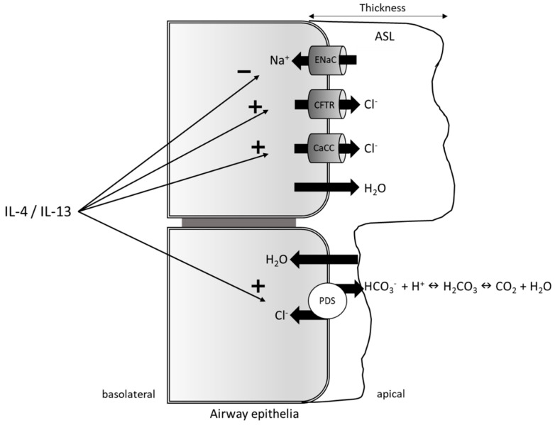
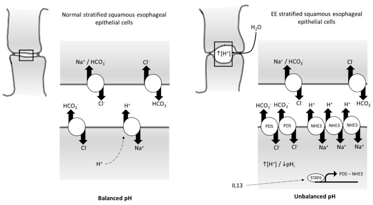
Pendrin (SLC26A4), a Cl-/anion exchanger, is expressed at high levels in kidney, thyroid, and inner ear epithelia, where it has an essential role in bicarbonate secretion/chloride reabsorption, iodide accumulation, and endolymph ion balance, respectively. Pendrin is expressed at lower levels in other tissues, such as airways and esophageal epithelia, where it is transcriptionally regulated by the inflammatory cytokines interleukin (IL)-4 and IL-13 through a signal transducer and activator of transcription 6 (STAT6)-mediated pathway. In the airway epithelium, increased pendrin expression during inflammatory diseases leads to imbalances in airway surface liquid thickness and mucin release, while, in the esophageal epithelium, dysregulated pendrin expression is supposed to impact the intracellular pH regulation system. In this review, we discuss some of the recent findings on interleukin-mediated transcriptional regulation of pendrin and how this dysregulation impacts airway and esophagus epithelial homeostasis during inflammatory diseases.
Keywords: airway epithelium; asthma; eosinophilic esophagitis; esophageal epithelium; interleukins; pendrin.
Conflict of interest statement
The authors declare no conflicts of interest.
Figures
Similar articles
-
Pendrin Mediates Bicarbonate Secretion and Enhances Cystic Fibrosis Transmembrane Conductance Regulator Function in Airway Surface Epithelia.Am J Respir Cell Mol Biol. 2019 Jun;60(6):705-716. doi: 10.1165/rcmb.2018-0158OC. Am J Respir Cell Mol Biol. 2019. PMID: 30742493
-
Transcriptional regulation of the pendrin gene.Cell Physiol Biochem. 2011;28(3):385-96. doi: 10.1159/000335100. Epub 2011 Nov 16. Cell Physiol Biochem. 2011. PMID: 22116353 Free PMC article. Review.
-
TNFα and IL-17 alkalinize airway surface liquid through CFTR and pendrin.Am J Physiol Cell Physiol. 2020 Aug 1;319(2):C331-C344. doi: 10.1152/ajpcell.00112.2020. Epub 2020 May 20. Am J Physiol Cell Physiol. 2020. PMID: 32432926 Free PMC article.
-
Pendrin function in airway epithelia.Cell Physiol Biochem. 2011;28(3):571-8. doi: 10.1159/000335115. Epub 2011 Nov 18. Cell Physiol Biochem. 2011. PMID: 22116372 Review.
-
Functional interplay between CFTR and pendrin: physiological and pathophysiological relevance.Front Biosci (Landmark Ed). 2022 Feb 21;27(2):75. doi: 10.31083/j.fbl2702075. Front Biosci (Landmark Ed). 2022. PMID: 35227018 Review.
Cited by
-
Chloride/Multiple Anion Exchanger SLC26A Family: Systemic Roles of SLC26A4 in Various Organs.Int J Mol Sci. 2024 Apr 10;25(8):4190. doi: 10.3390/ijms25084190. Int J Mol Sci. 2024. PMID: 38673775 Free PMC article. Review.
-
Overlap of Genomic and Transcriptomic Genes Identified in Familial Eosinophilic Esophagitis.Gastroenterology. 2025 Jun;168(6):1101-1113.e18. doi: 10.1053/j.gastro.2025.01.235. Epub 2025 Feb 4. Gastroenterology. 2025. PMID: 39914776
-
Multivariate gene expression-based survival predictor model in esophageal adenocarcinoma.Thorac Cancer. 2020 Oct;11(10):2896-2908. doi: 10.1111/1759-7714.13626. Epub 2020 Sep 1. Thorac Cancer. 2020. PMID: 32869505 Free PMC article.
-
Expression and Clinical Significance of Mucin Gene in Chronic Rhinosinusitis.Curr Allergy Asthma Rep. 2020 Aug 18;20(11):63. doi: 10.1007/s11882-020-00958-w. Curr Allergy Asthma Rep. 2020. PMID: 32812123 Free PMC article. Review.
-
Dynamic regulation of airway surface liquid pH by TMEM16A and SLC26A4 in cystic fibrosis nasal epithelia with rare mutations.Proc Natl Acad Sci U S A. 2023 Nov 21;120(47):e2307551120. doi: 10.1073/pnas.2307551120. Epub 2023 Nov 15. Proc Natl Acad Sci U S A. 2023. PMID: 37967223 Free PMC article.
References
-
- Royaux I.E., Suzuki K., Mori A., Katoh R., Everett L.A., Kohn L.D., Green E.D. Pendrin, the protein encoded by the Pendred syndrome gene (PDS), is an apical porter of iodide in the thyroid and is regulated by thyroglobulin in FRTL-5 cells. Endocrinology. 2000;141:839–845. doi: 10.1210/endo.141.2.7303. - DOI - PubMed
-
- Porra V., Bernier-Valentin F., Trouttet-Masson S., Berger-Dutrieux N., Peix J.L., Perrin A., Selmi-Ruby S., Rousset B. Characterization and semiquantitative analyses of pendrin expressed in normal and tumoral human thyroid tissues. J. Clin. Endocrinol. Metab. 2002;87:1700–1707. doi: 10.1210/jcem.87.4.8372. - DOI - PubMed
Publication types
MeSH terms
Substances
LinkOut - more resources
Full Text Sources
Research Materials
Miscellaneous
