. 2019 Mar;50(3):738-744.
doi: 10.1161/STROKEAHA.118.024058.

Rho Kinase Inhibition Blunts Lesion Development and Hemorrhage in Murine Models of Aggressive Pdcd10/Ccm3 Disease

Affiliations

Rho Kinase Inhibition Blunts Lesion Development and Hemorrhage in Murine Models of Aggressive Pdcd10/Ccm3 Disease

Robert Shenkar et al. Stroke. 2019 Mar.

Abstract

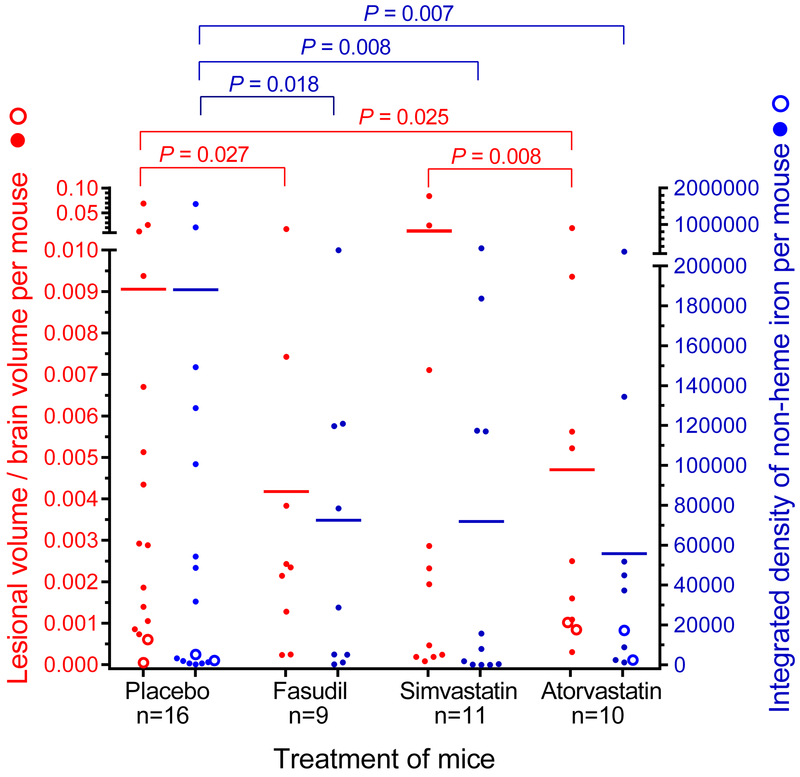
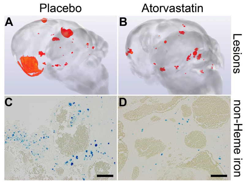
Background and Purpose- Previously, murine models Krit1 +/- Msh2 -/- and Ccm2 +/- Trp53 -/- showed a reduction or no effect on cerebral cavernous malformation (CCM) burden and favorable effects on lesional hemorrhage by the robust Rock (Rho-associated protein kinase) inhibitor fasudil and by simvastatin (a weak pleiotropic inhibitor of Rock). Herein, we concurrently investigated treatment of the more aggressive Pdcd10/Ccm3 model with fasudil, simvastatin, and higher dose atorvastatin to determined effectiveness of Rock inhibition. Methods- The murine models, Pdcd10 +/- Trp53 -/- and Pdcd10 +/- Msh2 -/-, were contemporaneously treated from weaning to 5 months of age with fasudil (100 mg/kg per day in drinking water, n=9), simvastatin (40 mg/kg per day in chow, n=11), atorvastatin (80 mg/kg per day in chow, n=10), or with placebo (n=16). We assessed CCM volume in mouse brains by microcomputed tomography. Lesion burden was calculated as lesion volume normalized to total brain volume. We analyzed chronic hemorrhage in CCM lesions by quantitative intensity of Perls staining in brain sections. Results- The Pdcd10 +/- Trp53 -/- /Msh2 -/- models showed a mean CCM lesion burden per mouse reduction from 0.0091 in placebos to 0.0042 ( P=0.027) by fasudil, and to 0.0047 ( P=0.025) by atorvastatin treatment, but was not changed significantly by simvastatin. Hemorrhage intensity per brain was commensurately decreased by Rock inhibition. Conclusions- These results support the exploration of proof of concept effect of high-dose atorvastatin on human CCM disease for potential therapeutic testing.

Keywords: atorvastatin; central nervous system; hemangioma; simvastatin; therapeutics.


Figures

Figure 1.
Figure 1.
Rock inhibition diminishes lesion burden (left axis, red) and lesion hemorrhage (right axis, blue) in combined Pdcd10+/−Trp53−/− (solid circles) and Pdcd10+/−Msh2−/− (hollow circles) models. Treatment with fasudil and atorvastatin, but not simvastatin, decreased lesion burden compared to contemporaneously raised placebos. Compared with placebos, treatment of mice with any of 3 Rock inhibitors decreased non-heme iron deposition in lesions. The 2-sided Conover 2-sample test was used to assess for significance.
Figure 2.
Figure 2.
Representative images generated from micro-computed tomography showing lesions (red) in brains (A, B) and from Perls staining of non-heme iron deposition (blue) depicting hemorrhage (C, D) in combined Pdcd10+/−Trp53−/− and Pdcd10+/−Msh2−/− models. Placebo mice had a greater lesion burden (A) and lesional hemorrhage (C) compared to those treated with atorvastatin (B, D). Scale bars are 100 μm.
Figure 3.
Figure 3.
Rock activity and inflammation within lesions of combined Pdcd10+/−Trp53−/− (solid circles) and Pdcd10+/−Msh2−/− (hollow circles) models treated by Rock inhibitors. (A) Rock inhibition did not affect the prevalence of lesion endothelial cells stained with phosphorylate myosin light chain (pMLC) compared with placebos. (B) The prevalence of lesion leukocytes stained with pMLC was decreased by 80% in mice treated with fasudil and was halved in those treated with atorvastatin compared with placebos. In contrast, simvastatin did not affect the prevalence of lesion leukocytes with Rock activity compared with placebos. (C) The total number of B lymphocytes per lesion was halved by simvastatin treatment, but unaffected by fasudil and atorvastatin, compared with placebos. (D) The total number of T lymphocytes per lesion was significantly decreased by fasudil, simvastatin and atorvastatin treatments compared with placebos. The number of lesions analyzed is designated by n, from 5 mice per group. The 2-sided Conover 2-sample test was used to assess for significant differences.
Figure 4.
Figure 4.
Attrition in combined Pdcd10+/−Trp53−/− and Pdcd10+/−Msh2−/− models. Kaplan-Meier plots show no significant effect of fasudil (n=9), simvastatin (n=13) or atorvastatin (n=17) treatment on survival compared with placebos (n=20) from weaning to the earliest age for the end of treatment (100 days of age). Although end of treatment ranged between 100 and 126 days of age, we compared survival curves up to 100 days, as all mice in the placebo and treatment groups were electively euthanized per intention to treat after at least 100 days. All animal attritions are reported and compared in Table IV in the online-only Data Supplement. There were neither overall significant differences (P=0.467), nor any individual significant differences between Rock inhibitor treatment and placebo groups (all P>0.24). When the entire 26-day range for ages of end of treatment were included in the analysis, there were neither overall significant differences (P=0.259), nor any individual significant differences between Rock inhibitor treatment and placebo groups (all P>0.10). The log-rank (Mantel-Cox) test was used to assess for significant differences.

Similar articles

Cited by

References

    1. Shenkar R, Shi C, Rebeiz T, Stockton RA, McDonald DA, Mikati AG, et al. Exceptional Aggressiveness of Cerebral Cavernous Malformation Disease Associated with Pdcd10 Mutations. Genet Med. 2015;17:188–196. - PMC - PubMed
    1. McDonald DA, Shenkar R, Shi C, Stockton RA, Akers AL, Kucherlapati MH, et al. A Novel Mouse Model of Cerebral Cavernous Malformations Based on the Two-Hit Mutation Hypothesis Recapitulates the Human Disease. Hum Mol Genet. 2011;20:211–222. - PMC - PubMed
    1. Gibson CC, Zhu W, Davis CT, Bowman-Kirigin JA, Chan AC, Ling J, et al. Strategy for Identifying Repurposed Drugs for the Treatment of Cerebral Cavernous Malformation. Circulation. 2015;131:289–299. - PMC - PubMed
    1. DiStefano PV, Kuebel JM, Sarelius IH, Glading AJ. Krit1 Protein Depletion Modifies Endothelial Cell Behavior Via Increased Vascular Endothelial Growth Factor (Vegf) Signaling. J Biol Chem. 2014;289:33054–33065. - PMC - PubMed
    1. Bravi L, Rudini N, Cuttano R, Giampietro C, Maddaluno L, Ferrarini L, et al. Sulindac Metabolites Decrease Cerebrovascular Malformations in Ccm3-Knockout Mice. Proc Natl Acad Sci U S A. 2015;112:8421–8426. - PMC - PubMed

Publication types

MeSH terms