. 2019 Feb 11;20(1):70.
doi: 10.1186/s12891-019-2452-0.

The commercial pig as a model of spontaneously-occurring osteoarthritis

Affiliations

The commercial pig as a model of spontaneously-occurring osteoarthritis

Mhairi A Macfadyen et al. BMC Musculoskelet Disord. .

Abstract

Background: Preclinical osteoarthritis models where damage occurs spontaneously may better reflect the initiation and development of human osteoarthritis. The aim was to assess the commercial pig as a model of spontaneous osteoarthritis development by examining pain-associated behaviour, joint cartilage integrity, as well as the use of porcine cartilage explants and isolated chondrocytes and osteoblasts for ex vivo and in vitro studies.

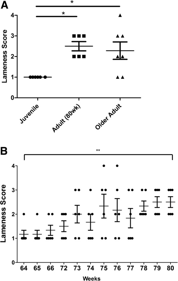
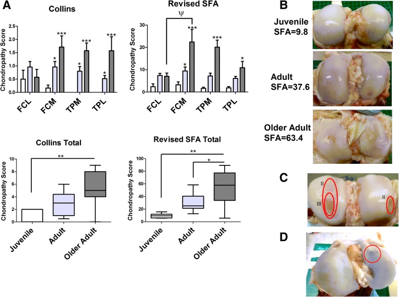
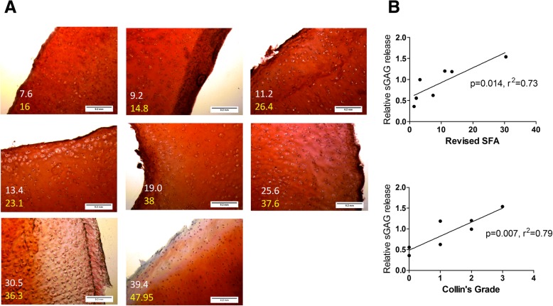
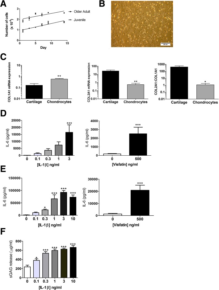
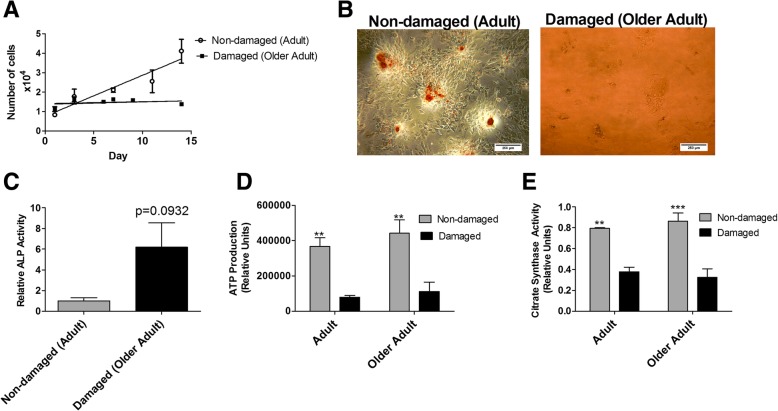
Methods: Female pigs (Large white x Landrace x Duroc) were examined at different ages from 6 weeks to 3-4 years old. Lameness was assessed as a marker of pain-associated behaviour. Femorotibial joint cartilage integrity was determined by chondropathy scoring and histological staining of proteoglycan. IL-6 production and proteoglycan degradation was assessed in cartilage explants and primary porcine chondrocytes by ELISA and DMMB assay. Primary porcine osteoblasts from damaged and non-damaged joints, as determined by chondropathy scoring, were assessed for mineralisation, proliferative and mitochondrial function as a marker of metabolic capacity.

Results: Pigs aged 80 weeks and older exhibited lameness. Osteoarthritic lesions in femoral condyle and tibial plateau cartilage were apparent from 40 weeks and increased in severity with age up to 3-4 years old. Cartilage from damaged joints exhibited proteoglycan loss, which positively correlated with chondropathy score. Stimulation of porcine cartilage explants and primary chondrocytes with either IL-1β or visfatin induced IL-6 production and proteoglycan degradation. Primary porcine osteoblasts from damaged joints exhibited reduced proliferative, mineralisation, and metabolic capacity.

Conclusion: In conclusion, the commercial pig represents an alternative model of spontaneous osteoarthritis and an excellent source of tissue for in vitro and ex vivo studies.

Keywords: Chondrocyte; Chondropathy; Osteoarthritis; Osteoblast; Pig.


Conflict of interest statement

Ethics approval and consent to participate

This study was approved by the University of Nottingham Animal Welfare Ethical Review Body (AWERB). The collection and use of human OA joint tissue was approved by the National Research Ethics Committee (NRES 13/NE/0222) and written informed consent was obtained from patients.

Consent for publication

Not applicable.

Competing interests

Dr. Simon Jones is a member of the Editorial Board of BMC Musculoskeletal Disorders. The authors declare that they have no other competing interests.

Publisher’s Note

Springer Nature remains neutral with regard to jurisdictional claims in published maps and institutional affiliations.

Figures

Fig. 1
Fig. 1
Development of lameness as a marker of behavioural pain. A scoring system was used to assess lameness as a marker of behavioural pain. a Comparison of median lameness score in juvenile (n = 6 animals), 80 wk. adult (n = 6 animals) and older adult (n = 7 animals) pigs. * = p < 0.05, significantly different from juvenile lameness score as determined by Kruskal-Wallis non-parametric test with Dunn’s post-hoc test. b Median lameness score in adult pigs across an 16 week timespan from age 64 to 80 wk. old (n = 6 animals). ** = p < 0.01, significant change in median score over time as determined by Kruskal-Wallis test
Fig. 2
Fig. 2
Spontaneous development of joint damage in the commercial pig. a Median total and joint compartment chondropathy score of femoral condyle and tibial plateau joints using Collin’s grading and Revised SFA in juvenile (white boxes, n = 6), adult (light grey boxes, n = 14) and older adult (dark grey boxes, n = 7) pigs. * = p < 0.05; ** = p < 0.01; *** = p < 0.001 significantly different compared to juvenile animals. ψ = p < 0.05 significantly different between medial and lateral compartment within same age group, as determined using Kruskal-Wallis non-parametric test. b Representative images of femoral condyle joints from juvenile, adult, and older adult animals. c Evidence of Grade II and Grade III cartilage lesions in femoral condyles of older adult pigs d Evidence of bony nodules in adult pigs. FCM = femoral condyle medial, FCL = femoral condyle lateral, TPM = tibital plateau medial, TPL = tibial plateau lateral
Fig. 3
Fig. 3
Areas of cartilage damage exhibit proteoglycan loss. a Representative images (10X magnification) of Safranin O staining of proteoglycan in femoral medial condyle cryosections from n = 8 adult pigs with varying signs of joint damage. White numbers represent SFA of the femoral condyle joint (medial plus lateral). Yellow numbers represent the SFA score of the whole joint (femoral condyle and tibial plateau). b Correlation between chondropathy scoring (SFA and Collins) and sGAG release from femoral condyle cartilage explants prepared from n = 7 adult pig joints. sGAG release was measured by DMMB assay and is expressed as the relative fold difference in damaged cartilage compared to healthy undamaged control cartilage explant. r = Pearson’s correlation coefficient
Fig. 4
Fig. 4
Characterisation of porcine chondrocytes and cartilage explant. a Proliferation of primary porcine chondrocytes isolated from juvenile (n = 6) and older adult pigs (n = 6). Proliferation was determined by MTS assay over a timecourse of 14 days. (B) Representative light microscope image (6.3X magnification) of porcine chondrocytes in 2D culture showing fibroblast-like morphology. c mRNA expression of type I and Type II collagen in primary porcine chondrocytes (n = 6 animals) compared to non-damaged porcine cartilage explant (n = 5 animals), from older adult pigs. Expression was determined by qRT-PCR normalised to total cDNA concentration. d Secretion of IL-6 from porcine primary chondrocytes from older adult pigs (n = 6) stimulated for 24 h with recombinant IL-1β (0.1–3 ng/ml) or recombinant visfatin (500 ng/ml). IL-6 in cell supernatants was measured by ELISA. * = p < 0.05; *** = p < 0.001 significantly different from un-stimulated control chondrocytes. Bars represent mean ± SEM (n = 6). e Secretion of IL-6 from porcine non-damaged cartilage explants from older adult pigs stimulated for 24 h with recombinant IL-1β (0.1–10 ng/ml) or recombinant visfatin (500 mg/ml) as measured by ELISA. * = p < 0.05; *** = p < 0.001 significantly different from un-stimulated control explants. Bars represent mean ± SEM (n = 20 explants per stimulant). f Detection of sulphated glycosaminoglycan (sGAG) proteoglycan side-chain upon 24 h stimulation of porcine non-damaged cartilage explant from older adult pigs with recombinant IL-1B. sGAG detected by DMMB assay. * = p < 0.05; *** = p < 0.001 significantly different from un-stimulated control explants. Bars represent mean ± SEM (n = 20 explants per stimulant)
Fig. 5
Fig. 5
a Proliferation of osteoblasts obtained from non-damaged adult and damaged older adult pig joints measured over a 14 day time period by MTS assay. Data points represent the mean cell number ± SEM (n = 3). b Representative light microscope images of alizarin red stained osteoblasts isolated from non-damaged adult and damaged older adult joints. c ALP activity isolated from non-damaged adult (n = 3 animals) and damaged older adult joints (n = 3 animals). Values represent mean ALP activity ± SEM. d Maximal mitochondrial ATP production in osteoblasts obtained from adult (n = 3 non-damaged; n = 3 damaged) and older adult (n = 3 non-damaged; n = 3 damaged) pig joints. ** = p < 0.01. Bars represent mean ± SEM. e Citrate synthase activity in osteoblasts obtained from adult (n = 3 non-damaged; n = 3 damaged) and older adult (n = 3 non-damaged; n = 3 damaged) pig joints. Bars represent mean ± SEM. ** = p < 0.01, *** = p < 0.001

References

    1. Murray CJ, Richards MA, Newton JN, Fenton KA, Anderson HR, Atkinson C, Bennett D, Bernabe E, Blencowe H, Bourne R, et al. UK health performance: findings of the global burden of disease study 2010. Lancet. 2013;381(9871):997–1020. doi: 10.1016/S0140-6736(13)60355-4. - DOI - PubMed
    1. Tonge DP, Pearson MJ, Jones SW. The hallmarks of osteoarthritis and the potential to develop personalised disease-modifying pharmacological therapeutics. Osteoarthr Cartil. 2014;22(5):609–621. doi: 10.1016/j.joca.2014.03.004. - DOI - PubMed
    1. Philp AM, Davis ET, Jones SW. Developing anti-inflammatory therapeutics for patients with osteoarthritis. Rheumatology (Oxford) 2017;56(6):869–881. - PubMed
    1. Chen A, Gupte C, Akhtar K, Smith P, Cobb J. The global economic cost of osteoarthritis: how the UK compares. Arthritis. 2012;2012:698709. doi: 10.1155/2012/698709. - DOI - PMC - PubMed
    1. Cross M, Smith E, Hoy D, Nolte S, Ackerman I, Fransen M, Bridgett L, Williams S, Guillemin F, Hill CL, et al. The global burden of hip and knee osteoarthritis: estimates from the global burden of disease 2010 study. Ann Rheum Dis. 2014;73(7):1323–1330. doi: 10.1136/annrheumdis-2013-204763. - DOI - PubMed

MeSH terms