. 2019 Feb 18;29(4):554-566.e4.
doi: 10.1016/j.cub.2019.01.009. Epub 2019 Feb 7.

Neural Substrates of Drosophila Larval Anemotaxis

Affiliations

Neural Substrates of Drosophila Larval Anemotaxis

Tihana Jovanic et al. Curr Biol. .

Abstract

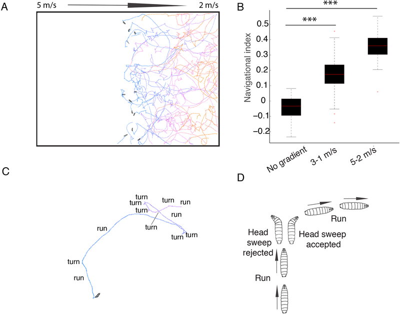
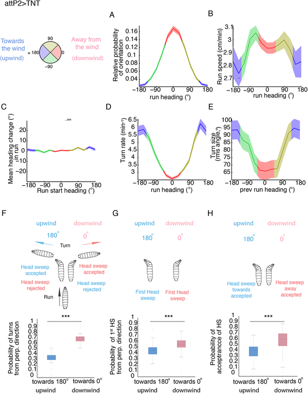
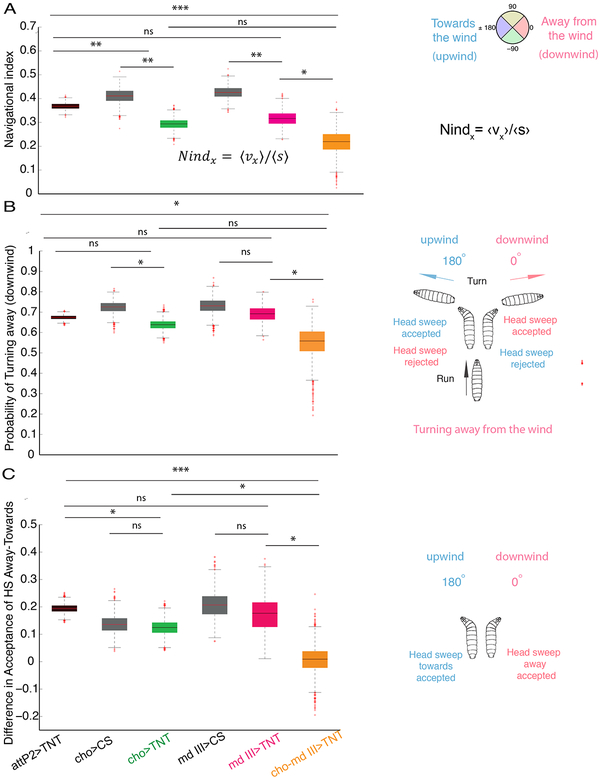
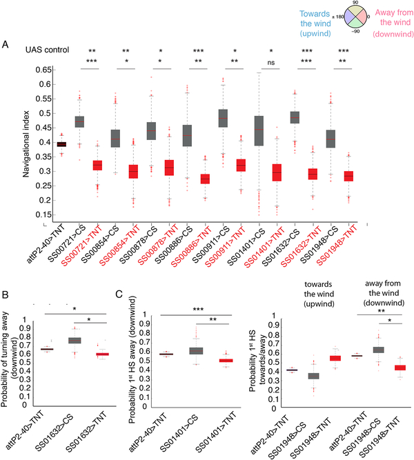
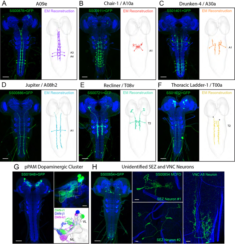
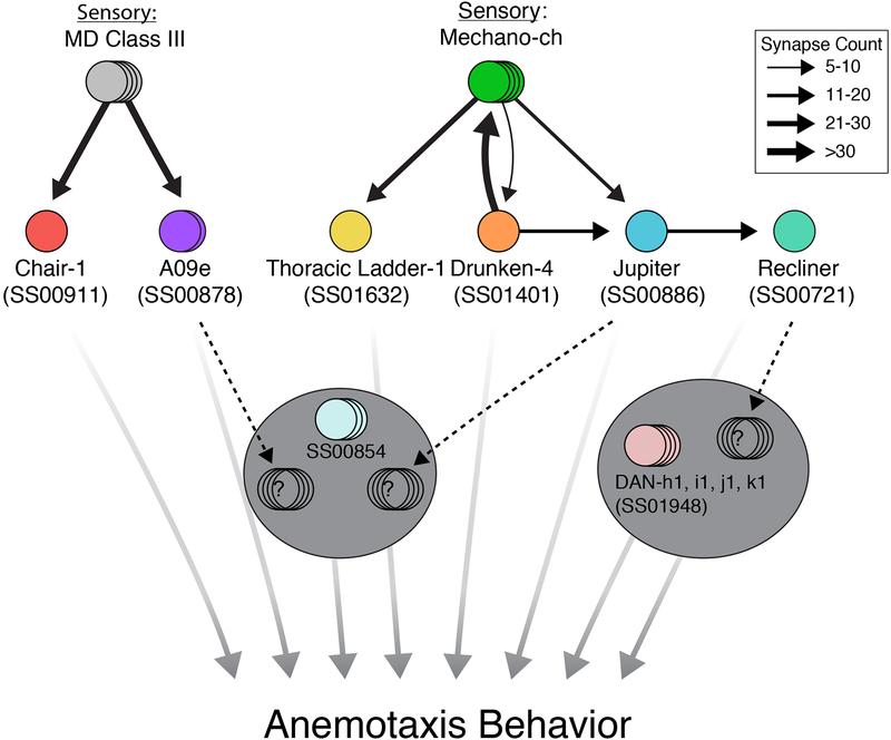
Animals use sensory information to move toward more favorable conditions. Drosophila larvae can move up or down gradients of odors (chemotax), light (phototax), and temperature (thermotax) by modulating the probability, direction, and size of turns based on sensory input. Whether larvae can anemotax in gradients of mechanosensory cues is unknown. Further, although many of the sensory neurons that mediate taxis have been described, the central circuits are not well understood. Here, we used high-throughput, quantitative behavioral assays to demonstrate Drosophila larvae anemotax in gradients of wind speeds and to characterize the behavioral strategies involved. We found that larvae modulate the probability, direction, and size of turns to move away from higher wind speeds. This suggests that similar central decision-making mechanisms underlie taxis in somatosensory and other sensory modalities. By silencing the activity of single or very few neuron types in a behavioral screen, we found two sensory (chordotonal and multidendritic class III) and six nerve cord neuron types involved in anemotaxis. We reconstructed the identified neurons in an electron microscopy volume that spans the entire larval nervous system and found they received direct input from the mechanosensory neurons or from each other. In this way, we identified local interneurons and first- and second-order subesophageal zone (SEZ) and brain projection neurons. Finally, silencing a dopaminergic brain neuron type impairs anemotaxis. These findings suggest that anemotaxis involves both nerve cord and brain circuits. The candidate neurons and circuitry identified in our study provide a basis for future detailed mechanistic understanding of the circuit principles of anemotaxis.

Keywords: CNS; Drosophila larva; anemotaxis; neural substrates; somatosensory processing.


Conflict of interest statement

Declaration of Interests

The authors declare no competing interest.

Figures

Figure 1.
Figure 1.. Drosophila Larvae perform negative anemotaxis.
A. In air-speed gradients, the larvae navigate down the gradient. The colors of the tracks represent the time from the beginning of the experiment (blue) to the end (orange). Snapshots of the initial positions of larvae in the center of the agar plate are shown B. Navigational index in 3–1 m/s and 5–2 m/s gradient compared to no-gradient conditions. In the box plot the median is indicated by the red horizontal line. Box boundaries represent first and third quartiles, whiskers extend to the most extreme data points not considered outliers. The outliers are indicated by red + C. Larvae alternate periods of runs and turns D. An example of a reorientation event where a larva perpendicular to the direction of the gradient accepts a head sweep and extends a run in a favorable direction is shown. P-values and N of animals are shown in Data S1 and Table S2, respectively).
Figure 2.
Figure 2.. Behavioral strategies of Drosophila larvae during anemotaxis.
Behavioral strategies in anemotaxis in control attP2>TNT (the same is shown for attP2-attP40>TNT in Figure S1). A compass in which 0° indicates the direction down the gradient (downwind) and 180° up the gradient (upwind) was used to keep track of larval direction during runs and turns as a function of the wind-speed spatial gradient. A. Relative probability of headings during runs. B. Speed versus heading during runs C. Mean heading change in runs D. Turn rate versus heading E. Turn size versus heading F. Distribution of turns from perpendicular direction G. Distribution of head sweeps from perpendicular direction H. Probability of starting a run during a head sweep A-E Values are mean and s.e.m. F-H. In the box plot the median is indicated by the horizontal line. Box boundaries represent first and third quartiles, whiskers extend to the most extreme data points not considered outliers. The outliers are indicated by red +. *: p<0.05, **: p<0.01, ***:
Figure 3.
Figure 3.. Somatosensory neurons implicated in anemotaxis.
A. Comparison of the navigation index in chordotonal>TNT (cho>TNT), multidendritic class III>TNT (md III>TNT), chordotonal-multidendritic class III>TNT (cho-md III>TNT) and control larvae. Silencing of cho and md III neurons simultaneously impairs anemotaxis. B. Probability of turns from perpendicular direction away from the wind (downwind) (0°) in cho>TNT, md III>TNT and cho-md III>TNT. Larvae with silenced cho and md III neurons together have a lower probability of turning away from the wind than the control and md III>TNT larvae C. Differences in probabilities of starting a run during a head sweep from perpendicular direction away and towards the wind. Larvae with silenced cho and md III neurons together have a lower difference in acceptance of head sweep away from and towards the wind compared to larvae with silenced md III and cho individually as well as control attP2>TNT larvae. Bootstrapped values are shown. In the box plot the median is indicated by the horizontal line. Box boundaries represent first and third quartiles, whiskers extend to the most extreme data points not considered outliers. The outliers are indicated by red +. *: p<0.05, **: p<0.01, ***:<p<0.001 (p-values can be found in Data S1 and S4, the N of animals in Table S2). See also Figure S2 and Data S2
Figure 4.
Figure 4.. Identifying central neurons whose silencing leads to reduced performance in anemotaxis.
A. Navigational indices of eight lines with less efficient anemotaxis compared to respective GAL4 controls and attP2-attP40>TNT control. Bootstrapped values are shown. *: p<0.05, **: p<0.01, ***:<p<0.001 (p-values can be found in Data S1). B. Probability of turns from perpendicular direction away from the wind (downwind) (0°) in SS01632>TNT compared to the controls attP2-attP40>TNT and SS01632>CS C. Probability of first head sweep from perpendicular direction away from the wind (downwind) (0°) in SS01401>TNT and SS01948>TNT compared to the control attP2-attP40>TNT Bootstrapped values are shown. In the box plot the median is indicated by the red horizontal line. Box boundaries represent first and third quartiles, whiskers extend to the most extreme data points not considered outliers. The outliers are indicated by red +. *: p<0.05, **: p<0.01, ***: <p<0.001 (p-values can be found in Data S1 and S4, the N of animals in Table S2). See also Figures S3, S4, Table S1 and Data S2
Figure 5.
Figure 5.. Electron microscopy correlates and connectivity of first-order anemotaxis neurons.
(A-F) Light microscopy z-projections of Split-GAL4s driving GFP expression in L3 (left panels) and the corresponding neuron reconstructed in a ssTEM L1 volume (right panels). A-C depict previously reconstructed neurons that have now been matched with Split-GAL4 lines. D-F depict neurons identified in EM and reconstructed for this study. Solid arrowheads indicate the ends of dendritic branches for comparison between light and EM images. Double arrowheads indicate contaminant neurons. (G) Expression pattern of SS01948 driving GFP expression in L3 (left panel). This line expresses in the pPAM cluster of four dopaminergic neurons that tile the mushroom body medial lobe (right panel). Note that four cell bodies are present, but only three DANs are presented in EM (DAN-h1 is not present in the ssTEM L1 volume and thus cannot be directly compared). (H) Expression pattern of SS00854 driving GFP expression in L3, depicting multiple neurons (left panel). MCFO revealed two SEZ neurons (middle panels) and an A8 VNC neuron (right panel). Scale bars are 50 μm unless indicated as 10 μm. See also Figure S5 and Data S5.
Figure 6.
Figure 6.. Anemotaxis circuit.
Chair-1 and A09e receive strong inputs from md III neurons, while Thoracic Ladder-1, Drunken-4, and Jupiter receive inputs from cho neurons. A second-order mechanosensory neuron, Recliner, was also identified downstream of Jupiter. Note that although no direct convergence between md III and cho neurons is reported here, two first-order neurons project to the SEZ (A09e and Jupiter) and the second-order Recliner projects to the brain at potential convergence sites (see dashed black arrows). No EM correlate has yet been found for SS00854. Width of black arrows indicates synaptic strength. All neurons reported here are involved in anemotaxis. See also Data S5

References

    1. Casas J, and Steinmann T (2014). Predator-induced flow disturbances alert prey, from the onset of an attack. Proc. Biol. Sci 281, 20141083–20141083. - PMC - PubMed
    1. Togunov RR, Derocher AE, and Lunn NJ (2017). Windscapes and olfactory foraging in a large carnivore. Sci. Rep 7, 46332. - PMC - PubMed
    1. Van Tilborg M, Sabelis MW, and Roessingh P (2004). State-dependent and odour-mediated anemotactic responses of the predatory mite Phytoseiulus persimilis in a wind tunnel. Exp. Appl. Acarol 32, 263–270. - PubMed
    1. Yu YSW, Graff MM, Bresee CS, Man YB, and Hartmann MJZ (2016). Whiskers aid anemotaxis in rats. Sci Adv 2, e1600716–e1600716. - PMC - PubMed
    1. Kalmus H (1942). Anemotaxis in Drosophila. Nature 150, 405–1.

Publication types

LinkOut - more resources