Review
. 2019 Sep 3;11(9):a034017.
doi: 10.1101/cshperspect.a034017.

Structure, Function, and Regulation of the Hsp90 Machinery

Affiliations
Review

Structure, Function, and Regulation of the Hsp90 Machinery

Maximilian M Biebl et al. Cold Spring Harb Perspect Biol. .

Abstract

Heat shock protein 90 (Hsp90) is a molecular chaperone involved in the maturation of a plethora of substrates ("clients"), including protein kinases, transcription factors, and E3 ubiquitin ligases, positioning Hsp90 as a central regulator of cellular proteostasis. Hsp90 undergoes large conformational changes during its ATPase cycle. The processing of clients by cytosolic Hsp90 is assisted by a cohort of cochaperones that affect client recruitment, Hsp90 ATPase function or conformational rearrangements in Hsp90. Because of the importance of Hsp90 in regulating central cellular pathways, strategies for the pharmacological inhibition of the Hsp90 machinery in diseases such as cancer and neurodegeneration are being developed. In this review, we summarize recent structural and mechanistic progress in defining the function of organelle-specific and cytosolic Hsp90, including the impact of individual cochaperones on the maturation of specific clients and complexes with clients as well as ways of exploiting Hsp90 as a drug target.


Figures

Figure 1.
Figure 1.
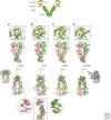
Structure of heat shock protein 90 (Hsp90) homologs. Structural models of (A) yeast Hsp90 (Protein Data Bank [PDB]: 2CG9), (B) mitochondrial tumor necrosis factor receptor associated protein 1 (TRAP1; PDB: 4IPE), (C) endoplasmic reticulum glucose-regulated protein 94 (Grp94; PDB: 5ULS), and (D) human cytosolic Hsp90β (PDB: 5FWL). For clarity, the Hsp90 protomers are distinctly colored. A schematic model of Hsp90 depicting the amino-terminal domain (NTD), middle domain (MD), and carboxy-terminal domain (CTD) as well as the linker and the adenosine triphosphate (ATP) lid is shown. In the top panel, the insets provide a zoomed view of the NTD of one protomer and the amino-terminal straps of the second protomer. In the bottom panel, the asymmetric conformation of the two protomers in TRAP1 is depicted (circles) and an alignment of the two protomers is shown (rectangle), highlighting the buckled conformation of the green protomer. Because the yeast Hsp82 and human Hsp90β structures were solved in complex with p23 (gray) or cell division control protein 37 (Cdc37; gray) and cyclin-dependent kinase 4 (Cdk4; blue), the full structures are shown on the left and right, respectively.
Figure 2.
Figure 2.
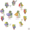
Hsp90 chaperone cycle. Hsp90 transitions through different conformational states during its ATPase cycle. Shown are intermediates of the cycle and how we envision client transfer and maturation. Some Hsp90 cochaperones preferentially bind specific Hsp90 conformations as indicated by the coloring of the circles in the middle and the association and dissociation of stress-inducible protein 1/Hsp70/Hsp90-organizing protein (Sti1/Hop) and large peptidylprolyl isomerases (PPIases) are depicted.
Figure 3.
Figure 3.
Hsp90 client comparison. Structural models showing the binding of Cdk4 (blue) to human Hsp90β and the glucocorticoid receptor (GR) ligand-binding domain (GR–LBD) (orange) to yeast Hsp82. The Hsp90β:Cdk4:Cdc37 complex is based on a cryogenic electron microscopy (cryo-EM) structure (PDB: 5FWL), whereas the Hsp90:GR–LBD:p23 structure is a pseudoatomic model combining data from cryo-EM, nuclear magnetic resonance (NMR) analysis, and small-angle X-ray scattering (SAXS) (Lorenz et al. 2014). Yeast Hsp82 (PDB: 2CG9) and human Hsp90β (PDB: 5FWL) were aligned in PyMOL to visualize the overlapping binding sites of the activator of Hsp90 ATPase protein 1 (Aha1) N-domain (red) and p23 (yellow) with the client-binding site. The binding site of the Aha1 N-domain was derived from an Aha1-N:Hsp90-MD crystal structure (PDB: 1USU) and docking experiments of Aha1-N on yeast Hsp82 (Retzlaff et al. 2010).

References

    1. Ai J, Wang Y, Dar JA, Liu J, Liu L, Nelson JB, Wang Z. 2009. HDAC6 regulates androgen receptor hypersensitivity and nuclear localization via modulating Hsp90 acetylation in castration-resistant prostate cancer. Mol Endocrinol 23: 1963–1972. 10.1210/me.2009-0188 - DOI - PMC - PubMed
    1. Åkerfelt M, Morimoto RI, Sistonen L. 2010. Heat shock factors: Integrators of cell stress, development and lifespan. Nat Rev Mol Cell Biol 11: 545–555. 10.1038/nrm2938 - DOI - PMC - PubMed
    1. Alexandrova EM, Yallowitz AR, Li D, Xu S, Schulz R, Proia DA, Lozano G, Dobbelstein M, Moll UM. 2015. Improving survival by exploiting tumour dependence on stabilized mutant p53 for treatment. Nature 523: 352–356. 10.1038/nature14430 - DOI - PMC - PubMed
    1. Ali MM, Roe SM, Vaughan CK, Meyer P, Panaretou B, Piper PW, Prodromou C, Pearl LH. 2006. Crystal structure of an Hsp90-nucleotide-p23/Sba1 closed chaperone complex. Nature 440: 1013–1017. 10.1038/nature04716 - DOI - PMC - PubMed
    1. Altieri DC, Stein GS, Lian JB, Languino LR. 2012. TRAP-1, the mitochondrial Hsp90. Biochim Biophys Acta 1823: 767–773. 10.1016/j.bbamcr.2011.08.007 - DOI - PMC - PubMed

Publication types

LinkOut - more resources