. 2019 Mar 4;216(3):571-586.
doi: 10.1084/jem.20181589. Epub 2019 Feb 11.

Chronic virus infection compromises memory bystander T cell function in an IL-6/STAT1-dependent manner

Affiliations

Chronic virus infection compromises memory bystander T cell function in an IL-6/STAT1-dependent manner

Isabel Barnstorf et al. J Exp Med. .

Abstract

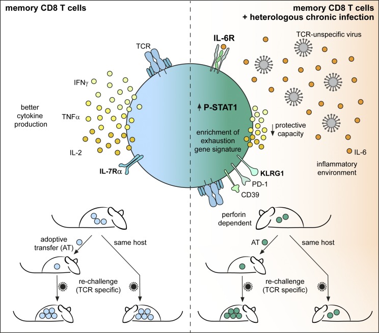
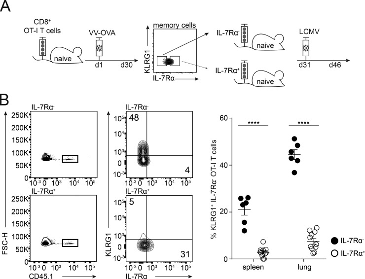
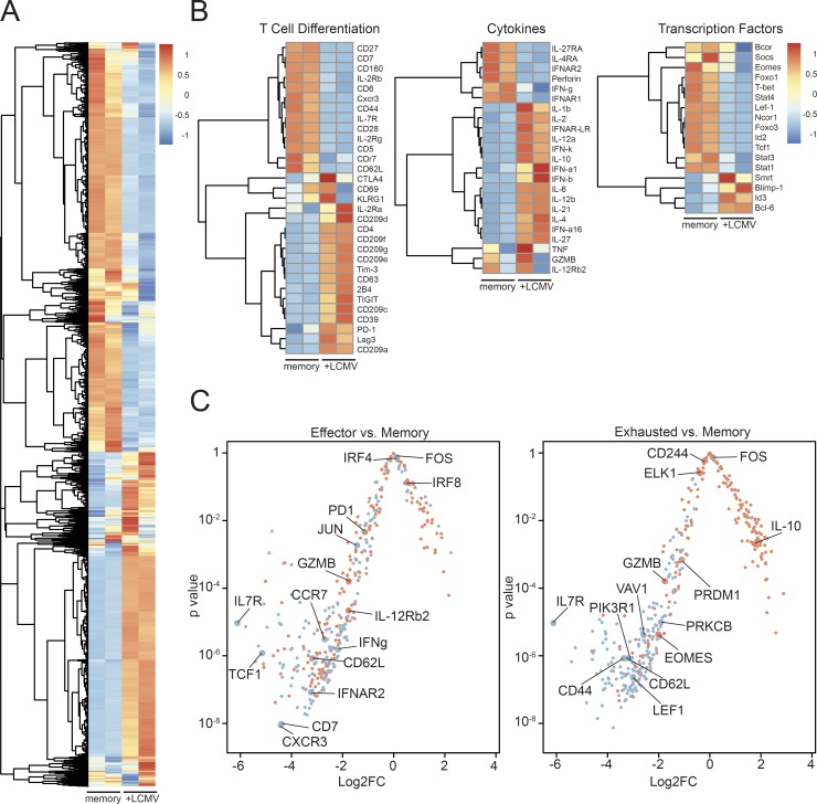
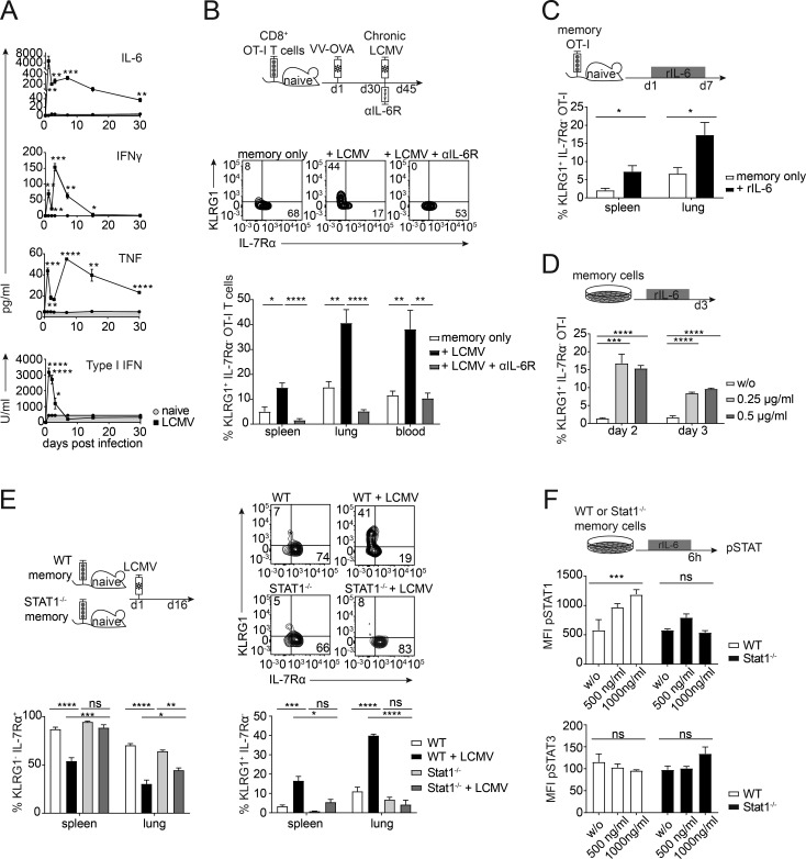
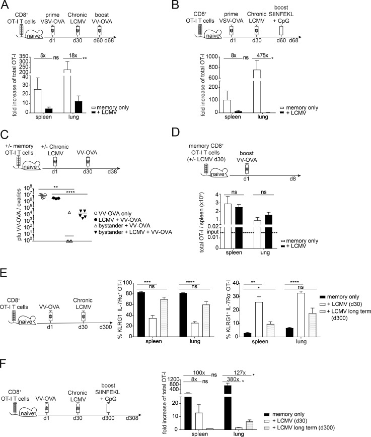
Chronic viral infections are widespread among humans, with ∼8-12 chronic viral infections per individual, and there is epidemiological proof that these impair heterologous immunity. We studied the impact of chronic LCMV infection on the phenotype and function of memory bystander CD8+ T cells. Active chronic LCMV infection had a profound effect on total numbers, phenotype, and function of memory bystander T cells in mice. The phenotypic changes included up-regulation of markers commonly associated with effector and exhausted cells and were induced by IL-6 in a STAT1-dependent manner in the context of chronic virus infection. Furthermore, bystander CD8 T cell functions were reduced with respect to their ability to produce inflammatory cytokines and to undergo secondary expansion upon cognate antigen challenge with major cell-extrinsic contributions responsible for the diminished memory potential of bystander CD8+ T cells. These findings open new perspectives for immunity and vaccination during chronic viral infections.


Figures

None
Graphical abstract
Figure 1.
Figure 1.
Chronic virus infection significantly reduces total numbers of memory bystander T cells. (A) Experimental approach. Adoptive transfer of CD45.1+ TCR-transgenic OVA257–264-specific (OT-I) CD8+ T cells into congenic CD45.2 WT C57BL/6 mice, followed by priming with VV-OVA. On day 30 after priming, mice were infected with the persistent docile strain of LCMV with an intermediate (104 ffu) or high (106 ffu) dose. The experiment was terminated 30 d after LCMV infection. (B) Viral titers in liver and kidney at day 30 after infection. (C) Total cell counts of OT-I memory bystander CD8+ T cells in spleen, lung, and blood of memory only (white, no LCMV infection), memory + LCMV intermediate dose (gray), and memory + LCMV high-dose (black) 30 d after infection. (B and C) One representative experiment out of two is shown with four to five mice per group. Statistical analysis was performed using the unpaired two-tailed Student’s t test: *, P < 0.05; ***, P < 0.001; ****, P < 0.0001. Error bars show SEM.
Figure 2.
Figure 2.
Loss of memory bystander T cells partially due to perforin-mediated cytotoxicity. (A) Experimental approach. Adoptive transfer of CD45.1+ OT-I memory CD8+ T cells (≥30 d after VV-OVA infection) into congenic CD45.2 WT C57BL/6 or PKOB−/− mice. Mice were infected with LCMV docile 1 d after transfer. The experiment was terminated 10 d after LCMV infection. (B) Top: Total cell counts of OT-I memory bystander CD8+ T cells in spleen and lung of memory only in WT (black, uninfected), memory + LCMV in WT (white), memory only in PKOB−/− (dark gray, uninfected), and memory + LCMV in PKOB−/− (light gray). Bottom: Quantification of IL-7Rα+ OT-I T cell percentages in spleen and lung of memory only in WT (black, uninfected), memory + LCMV in WT (white), memory only in PKOB−/− (dark gray, uninfected), and memory + LCMV in PKOB−/− (light gray). One representative experiment out of two with four mice per group is shown. Statistical analysis was performed using the unpaired two-tailed Student’s t test: ns, P ≥ 0.05; **, P < 0.01; ****, P < 0.0001. (C) Immunofluorescence staining of splenic thin sections of WT (top row), WT+LCMV (second row from top), PKOB−/− (third row from top), or PKOB−/− + LCMV (bottom row) for B cell zone (B220+, red), T cell zone (podoplanin, yellow), and T cells (CD3+, white). Scale bar, 90 µm. ROI, dashed white line. (D) Top: Ratio of volume of CD3 to volume of podoplanin in the ROI. 20 ROIs were analyzed per group. Bottom: Ratio of volume of podoplanin per ROI. Twenty ROIs were analyzed per group. Error bars show SEM.
Figure 3.
Figure 3.
Chronic virus infection skews memory bystander phenotype and function. (A) Experimental approach. Adoptive transfer of CD45.1+ OT-I CD8+ T cells into congenic CD45.2 WT C57BL/6 mice, followed by priming with VV-OVA. On at least day 30 after priming, the mice were infected with LCMV docile at an intermediate (104 ffu) or high (106 ffu) dose. The experiment was terminated 30 d after LCMV infection. (B) Memory bystander CD8+ T cells in the spleen, lung, and blood. Flow cytometry plots pregated on live CD8+ T cells from spleen are depicted. Representative FACS plots are shown for CD45.1+ bystander T cells (memory only, + LCMV intermediate, and + LCMV high) and their expression of KLRG1 and IL-7Rα. Right: Quantification of percentages of KLRG1+ IL-7Rα OT-I T cells and KLRG1 IL-7Rα+ OT-I T cells shown for memory only (white, uninfected), + LCMV intermediate dose (gray), and + LCMV high dose (black) in the spleen and lung. FSC-H, forward scatter height. (C) Representative flow cytometry plots of cytokine production and quantification of IFNγ, IL-2, and TNFα production by memory bystanders from memory only (white, uninfected), + LCMV intermediate dose (gray), and + LCMV high dose (black) in the lung after 6 h of restimulation with the cognate antigen. (D) Left: Experimental approach. Mice were infected with a high-dose LCMV docile. 30 d after infection, memory bystander CD8+ T cells were adoptively transferred into chronically infected mice. Bystander T cells were analyzed 30 d after transfer. Right: Quantification of percentages of KLRG1+ IL-7Rα OT-I T cells and KLRG1 IL-7Rα+ OT-I T cells shown for memory only (white, uninfected) and + LCMV (black) in the spleen and lung. (B–D) One representative experiment out of two is shown with four mice per group. Statistical analysis was performed using the unpaired two-tailed Student’s t test: ns, P ≥ 0.05; *, P < 0.05; **, P < 0.01; ***, P < 0.001; ****, P < 0.0001. Error bars show SEM.
Figure 4.
Figure 4.
KLRG1+ population arises from the IL-7Rαlow compartment. (A) Experimental approach. Adoptive transfer of CD45.1+ OT-I CD8+ T cells into congenic CD45.2 WT C57BL/6 mice, followed by priming with VV-OVA. On day 30 after priming, OT-I memory cells were sorted for KLRG1 IL-7Rαlow and KLRG1 IL-7Rαhigh and adoptively transferred into age- and sex-matched naive mice. 1 d after transfer, mice were chronically infected with LCMV docile. The experiment was terminated 15 d after LCMV infection. (B) Memory bystander CD8+ T cells in spleen and lung. Left: Flow cytometry plots pregated on live CD8+ T cells are shown. Representative FACS plots for CD45.1+ memory only and memory + LCMV and their expression of KLRG1 and IL-7Rα. Right: Quantification of percentages of KLRG1+ IL-7Rα OT-I T cells are shown for IL-7Rα+ (white) and IL-7Rα (black) in the spleen and lung. FSC-H, forward scatter height. One representative experiment out of two is shown with five mice per group. Statistical analysis was performed using the unpaired two-tailed Student’s t test: *, P < 0.05; ****, P < 0.0001. Error bars show SEM.
Figure 5.
Figure 5.
Transcriptional profile of memory bystander CD8+ T cells induced by chronic virus infection. (A) Heat map of differentially expressed genes comparing memory only (memory, uninfected) and memory + LCMV (+LCMV) infection. (B) Heat maps for selected genes of interest separated into categories of T cell differentiation, cytokines, and transcription factors comparing memory only (memory, uninfected) and memory + LCMV (+LCMV) infection. (C) Log2 fold change (Log2FC) and accompanying P values are shown for genes reported in four gene sets (Wherry et al., 2007). Red points correspond to those genes reported as up-regulated in the effector T cell gene set (left) or exhausted T cell gene set (right), whereas blue points correspond to those genes reported as down-regulated in the effector T cell gene set (left) or exhausted T cell gene set (right).
Figure 6.
Figure 6.
IL-6 drives phenotypic alterations of memory bystander T cells. (A) Cytokine levels in the serum over 30 d of infection (LCMV, open squares) and naive controls (naive, gray circles, gray area) were measured by CBA (IL-6, IFNγ, and TNF) or IFN bioassay (type I IFNs). (B) Top: Experimental approach. Adoptive transfer of CD45.1+ OT-I CD8+ T cells into congenic CD45.2 WT C57BL/6 mice, followed by priming with VV-OVA. On day 30 after priming, mice were persistently infected with LCMV docile. From the onset of the infection, αIL-6R blocking antibody was injected i.p. every second day. The experiment was terminated 15 d after LCMV infection. Middle: Representative flow cytometry plots of the expression of KLRG1 and IL-7Rα of memory only, + LCMV, and +LCMV + αIL-6R. Bottom: Quantification of percentages of KLRG1+ IL-7Rα OT-I T cells shown for memory only (white, uninfected) + LCMV (black), and +LCMV + αIL-6R (gray) in the spleen, lung, and blood. (C) Top: Experimental approach. Adoptive transfer of CD45.1+ OT-I CD8+ T cells into congenic CD45.2 WT C57BL/6 mice, followed by priming with VV-OVA. On day 30 after priming, mice were injected with 1 µg of rIL-6 every day for 1 wk. Bottom: Quantification of percentages of KLRG1+ IL-7Rα OT-I T cells shown for memory only (white, uninfected) and +rIL-6 (black) in the spleen and lung. (D) Top: Experimental approach. FACS-sorted memory OT-I CD8+ T cells were cultured in vitro with or without rIL-6 for 3 d. Bottom: Quantification of percentages of KLRG1+ OT-I T cells shown for no cytokine (w/o, white), 0.25 µg/ml (light gray), and 0.5 µg/ml (dark gray). (E) Top left: Experimental approach. Adoptive transfer of 5 × 10⁵ CD45.1+ WT memory OT-I or CD45.1+ Stat1−/− memory OT-I CD8+ T cells into congenic CD45.2+ WT C57BL/6 mice. 1 d after transfer, mice were persistently infected with LCMV docile. The experiment was terminated 15 d after LCMV infection. Top right: Representative flow cytometry plots of the expression of KLRG1 and IL-7Rα for WT OT-I ± LCMV and Stat1−/− OT-I ± LCMV memory bystander T cells. Bottom: Quantification of percentages of KLRG1+ IL-7Rα OT-I T cells and KLRG1 IL-7Rα+ OT-I T cells shown for WT (white, uninfected), WT + LCMV (black), Stat1−/− (light gray), and Stat1−/− + LCMV (dark gray) in the spleen and lung. (F) Experimental approach. FACS-sorted WT or Stat1−/− memory OT-I CD8 T cells were cultured in vitro with or without rIL-6 for 6 h. Bottom: Quantification of mean fluorescence intensity (MFI) of pSTAT1+ and pSTAT3+ WT (white) or Stat1−/− (black) OT-I T cells shown for no cytokine (w/o), 500 ng/ml and 1000 ng/ml. One representative experiment out of two is shown with four to five mice per group (A–C and E) or triplicates for the in vitro experiments (D and F). Statistical analysis was performed using the unpaired two-tailed Student’s t test: ns, P ≥ 0.05; *, P < 0.05; **, P < 0.01; ***, P < 0.001; ****, P < 0.0001. Error bars show SEM.
Figure 7.
Figure 7.
Chronic viral infection negatively impacts secondary expansion and protection of memory bystanders in a cell-extrinsic manner. (A) Top: Experimental approach. Adoptive transfer of 3 × 10⁵ CD45.1+ OT-I CD8+ T cells into congenic CD45.2 WT C57BL/6 mice, followed by priming with VSV-OVA. On day 30 after priming, mice were persistently infected with LCMV docile. 30 d after LCMV infection mice were infected with VV-OVA. Memory bystanders were analyzed for expansion 8 d after VV-OVA challenge. Bottom: Quantification of fold increase of OT-I T cells shown for memory only (white, uninfected) and + LCMV (black) in the spleen and in the lung. (B) Top: Experimental approach. Adoptive transfer of CD45.1+ OT-I CD8+ T cells into congenic CD45.2 WT C57BL/6 mice, followed by priming with VV-OVA. On day 30 after priming, mice were persistently infected with LCMV docile. 30 d after LCMV infection a vaccination boost of 50 µg SIINFEKL peptide and 10 µg CpG was injected intravenously. Memory bystanders were analyzed for expansion 8 d after challenge. Bottom: Quantification of fold increase of OT-I T cells shown for memory only (white, uninfected) and + LCMV (black) in the spleen and in the lung. (C) Top: Experimental approach. Adoptive transfer of 5 × 10⁵ CD45.1+ OT-I memory CD8+ T cells into congenic CD45.2 WT C57BL/6 mice. 1 d after transfer, mice were persistently infected with LCMV docile; controls remained uninfected. 30 d after LCMV infection, mice were injected intravenously with VV-OVA. Further controls were WT C57BL/6 mice that were persistently infected with LCMV docile or were left uninfected as controls. VV-OVA titers were analyzed 8 d after VV-OVA infection in the ovaries. Bottom: Quantification of PFUs VV-OVA/ovaries shown for VV-OVA only (circles, white), LCMV + VV-OVA (circles, black), bystander + VV-OVA (triangle, white), and bystander + LCMV + VV-OVA (triangle, black). (D) Top: Experimental approach. Adoptive transfer of 103 CD45.1+ OT-I memory CD8+ T cells, which had or had not been exposed to chronic LCMV infection for 30 d, into congenic CD45.2 WT C57BL/6 mice. 1 d after transfer, mice were infected with VV-OVA. Memory bystanders were analyzed for expansion 8 d after challenge. Bottom: Quantification of total numbers of OT-I T cells shown for memory only (white, uninfected) and + LCMV (black) in the spleen and in the lung. (E) Left: Experimental approach. Adoptive transfer of CD45.1+ OT-I CD8+ T cells into congenic CD45.2 WT C57BL/6 mice, followed by priming with VV-OVA. On day 30 after priming, mice were persistently infected with LCMV docile. The experiment was terminated after viral clearance at day 300. Right: Quantification of percentages of KLRG1+ IL-7Rα and KLRG1 IL-7Rα+ OT-I T cells shown for memory only (black, uninfected), + LCMV (day 30, white), and +LCMV long term (day 300, white with black dots) in the spleen and in the lung. (F) Left: Experimental approach. Adoptive transfer of CD45.1+ OT-I CD8+ T cells into congenic CD45.2 WT C57BL/6 mice, followed by priming with VV-OVA. On day 30 after priming, mice were persistently infected with LCMV docile. After viral clearance at day 300, a vaccination boost of 50 µg SIINFEKL peptide and 10 µg CpG was injected intravenously. As a control, day 30 LCMV mice were included. Memory bystanders were analyzed for expansion 8 d after challenge. Right: Quantification of fold increase of total OT-I T cells shown for memory only (black, uninfected), + LCMV (day 30, white), and + LCMV long-term (day 300, white with black dots) in the spleen and in the lung. (A–F) One representative experiment out of two is shown with three to five mice per group. Statistical analysis was performed using the unpaired two-tailed Student’s t test: ns, P ≥ 0.05; *, P < 0.05; **, P < 0.01; ***, P < 0.001; ****, P < 0.0001. Error bars show SEM.

Comment in

References

    1. Bahl K., Kim S.K., Calcagno C., Ghersi D., Puzone R., Celada F., Selin L.K., and Welsh R.M.. 2006. IFN-induced attrition of CD8 T cells in the presence or absence of cognate antigen during the early stages of viral infections. J. Immunol. 176:4284–4295. 10.4049/jimmunol.176.7.4284 - DOI - PubMed
    1. Bastidas S., Graw F., Smith M.Z., Kuster H., Günthard H.F., and Oxenius A.. 2014. CD8+ T cells are activated in an antigen-independent manner in HIV-infected individuals. J. Immunol. 192:1732–1744. 10.4049/jimmunol.1302027 - DOI - PubMed
    1. Battegay M., Cooper S., Althage A., Bänziger J., Hengartner H., and Zinkernagel R.M.. 1991. Quantification of lymphocytic choriomeningitis virus with an immunological focus assay in 24- or 96-well plates. J. Virol. Methods. 33:191–198. 10.1016/0166-0934(91)90018-U - DOI - PubMed
    1. Beura L.K., Hamilton S.E., Bi K., Schenkel J.M., Odumade O.A., Casey K.A., Thompson E.A., Fraser K.A., Rosato P.C., Filali-Mouhim A., et al. . 2016. Normalizing the environment recapitulates adult human immune traits in laboratory mice. Nature. 532:512–516. 10.1038/nature17655 - DOI - PMC - PubMed
    1. Brune W., Hengel H., and Koszinowski U.H.. 2001. A mouse model for cytomegalovirus infection. Curr. Protoc. Immunol. Chapter 19:7 10.1002/0471142735.im1907s43 - DOI - PubMed

Publication types

MeSH terms