Review
. 2019 May;20(5):303-320.
doi: 10.1038/s41580-019-0103-9.

Haematopoietic stem cell activity and interactions with the niche

Affiliations
Review

Haematopoietic stem cell activity and interactions with the niche

Sandra Pinho et al. Nat Rev Mol Cell Biol. 2019 May.

Abstract

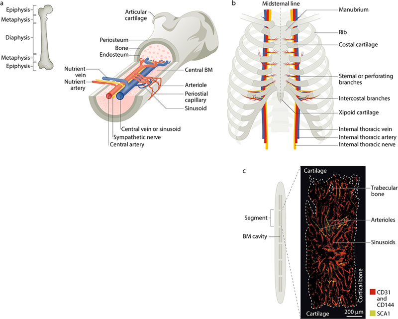
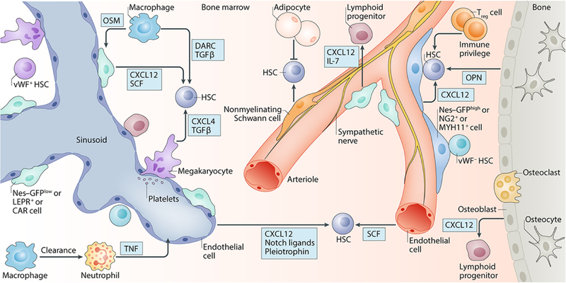
The haematopoietic stem cell (HSC) microenvironment in the bone marrow, termed the niche, ensures haematopoietic homeostasis by controlling the proliferation, self-renewal, differentiation and migration of HSCs and progenitor cells at steady state and in response to emergencies and injury. Improved methods for HSC isolation, driven by advances in single-cell and molecular technologies, have led to a better understanding of their behaviour, heterogeneity and lineage fate and of the niche cells and signals that regulate their function. Niche regulatory signals can be in the form of cell-bound or secreted factors and other local physical cues. A combination of technological advances in bone marrow imaging and genetic manipulation of crucial regulatory factors has enabled the identification of several candidate cell types regulating the niche, including both non-haematopoietic (for example, perivascular mesenchymal stem and endothelial cells) and HSC-derived (for example, megakaryocytes, macrophages and regulatory T cells), with better topographical understanding of HSC localization in the bone marrow. Here, we review advances in our understanding of HSC regulation by niches during homeostasis, ageing and cancer, and we discuss their implications for the development of therapies to rejuvenate aged HSCs or niches or to disrupt self-reinforcing malignant niches.


Conflict of interest statement

Competing interests

The authors declare no competing interests.

Figures

Figure 1.
Figure 1.. Microarchitecture of the adult mouse femur and sternum bone marrow.
Postnatally, the bone marrow (BM) is the primary site of haematopoietic stem cell (HSC) maintenance and haematopoiesis. a | Longitudinal view of a femur illustrating the arrangement of blood vessels and nerves (marked in yellow) that accompany the arteries within the BM cavity. The periosteal layer covers the outer surface of the bone and the endosteal layer is at the interface of bone and BM. Branching arteries (red) run parallel to the long axis of the marrow cavity, often close to the endosteum. These vessels feed into the sinusoidal network, which is distributed evenly throughout the marrow cavity and then coalesces to form the venous circulation (blue). b | Anterior view of the adult human sternum with attached ribs. Unlike the long bones, which mostly contain adipocytes in adult humans, the sternum has rich haematopoietic activity in both mice and humans, which makes it a suitable site to study haematopoiesis. The blood supply to the sternum originates from sternal/perforating branches located in the intersegmental spaces (between the ribs) from internal thoracic arteries that extend along the inside of the ribcage parallel to the sternum. c | Mid-sagittal section of the mouse sternum illustrating the six BM compartments (the human sternum comprises two bones, the manubrium and the body) with a representative image of a sternal segment showing the vasculature, which is labelled using intravenously injected antibodies against CD31/CD144 (red) and SCA1 (green). Arterioles can be distinguished from the sinusoidal network by their SCA1high expression.
Figure 2.
Figure 2.. Cellular and molecular constituents of the HSC niche.
Various cell types have been implicated in regulating haematopoietic stem cell (HSC) activity, including perivascular mesenchymal stem cells (MSCs), endothelial cells, osteoblasts, adipolineage cells, sympathetic nervous system (SNS) nerves, nonmyelinating Schwann cells, macrophages, megakaryocytes and regulatory T (Treg) cells. The target plot illustrates how bone marrow (BM) niche cells contribute to HSC regulation indirectly or directly by synthesizing niche factors in the form of cell-bound or secreted molecules. The colour of the radial spokes indicates the HSC activity that is affected. *Molecules involved in bone marrow regeneration after ablation. Bold type indicates those molecules for which there is functional data using cell-specific genetic evidence. SCF, stem cell factor; CXCL12, CXC-chemokine ligand 12; CXCL4, CXC chemokine ligand 4; FGF1, fibroblast growth factor 1; TGFβ, transforming growth factor-β; DARC, duffy antigen receptor for chemokines; GP130, glycoprotein 130; VCAM1, vascular cell adhesion molecule 1; IL-10, interleukin-10.
Figure 3.
Figure 3.. The adult bone marrow HSC niche in homeostasis.
Schematic representation of the adult bone marrow haematopoietic stem cell (HSC) niche in homeostasis, showing various cell types and niche factors that directly or indirectly regulate HSC activity. Emerging evidence highlights the vasculature and associated stromal cells, such as periarteriolar Nes–GFPhigh cells, NG2+ cells and MYH11+ cells and perisinusoidal Nes–GFPlow cells, CAR cells and LEPR+ cells, as key regulators of HSC maintenance. Sympathetic nervous system (SNS) nerves regulate HSC mobilization and non-myelinating Schwann cells may contribute to HSC quiescence. Osteoblasts have been implicated in HSC regulation but the precise molecular signals have not been clearly elucidated; however, they may have a role in regulating lymphoid progenitors. Adipocytes may negatively affect HSC maintenance. Haematopoietic cells, such as macrophages, neutrophils, regulatory T (Treg) cells and megakaryocytes, are examples of HSC-derived progeny that can feedback and contribute to HSC maintenance or mobilization. Regional localization of HSC subsets has shown that platelet-biased or myeloid-biased Vwf–GFP+ HSCs and Vwf–GFP HSCs are located in, and regulated by, separate BM niches containing megakaryocytes and arterioles, respectively. CAR cells, CXCL12-abundant reticular cells; SCF, stem cell factor; CXCL4, C-X-C-chemokine ligand 4; CXCL12, CXC chemokine ligand 12; DARC, duffy antigen receptor for chemokines; IL-7, interleukin-7; LEPR, leptin receptor; MYH11, myosin heavy chain 11; NG2, neuron/glial antigen 2; OPN, osteopontin; OSM, oncostatin M; SCF, stem cell factor; TGFβ, transforming growth factor-b; TNF, tumour necrosis factor; vWF, von Willebrand factor.
Figure 4.
Figure 4.. The adult bone marrow HSC niche in ageing and malignancy.
a | Schematic representation of the aged haematopoietic stem cell (HSC) niche. Aged HSCs are highly proliferative and exhibit increased myeloid-biased differentiation and reduced regenerative capacity. Ageing-related alterations of the bone marrow (BM) niche that influence HSC ageing include alterations in the vasculature and mesenchymal stem and progenitor cells (MSPCs), increased adipogenesis and reduced osteogenesis, altered secretion of niche factors and a reduced number of adrenergic nerves. b | HSC niche alterations that promote cancer. Epigenetic or genetic lesions (lightning bolt) in stromal niche regulators can lead to the loss of inhibitory signals that control the growth of premalignant clones and ultimately promote myeloid malignancies. These lesions include alterations in the expression of RBPJ in endothelial cells, β-catenin in osteoblasts and Dicer 1, SBDS and PTPN11 in MSPCs; and deletion of Rarg (which encodes RARγ), Rb or Mib1 in undefined stromal cells. c | Cancer promotes niche remodelling. The majority of myeloid malignancies are caused by epigenetic and/or genetic mutations in haematopoietic stem and progenitor cells (HSPCs), which lead to BM niche remodelling that supports cancer cell growth at the expense of normal haematopoiesis. Alterations produced by different malignancies can lead to a proinflammatory environment characterized by impaired MSPC differentiation, fibrosis, vascular remodelling, neuropathy and reduced production of HSC-niche factors by stromal cells. Leukemic stem cells (LSCs) can also upregulate the expression of CXCR4, VLA4 and CD44, to hijack the mechanisms of adhesion used by healthy HSPCs. ADRβ3, adrenergic β3 receptor; AML, acute myeloid leukaemia; CCL3, CC chemokine ligand 3; CML, chronic myeloid leukaemia; CXCL12, CXC chemokine ligand 12; CXCR4, CXC chemokine receptor type 4; IL-1 β, interleukin-1β; MDS, myelodysplastic syndrome; MIB1, mind bomb 1; MPN, myeloproliferative neoplasm; NG2, neuron/glial antigen 2; OPN, osteopontin; PTPN11, protein tyrosine phosphatase, non-receptor type 11; RARγ, retinoic acid receptor-γ; RB, retinoblastoma protein; RBPJ, recombination signal binding protein for immunoglobulin-κ J region; ROS, reactive oxygen species; SBDS, Shwachman–Bodian–Diamond syndrome protein; SCF, stem cell factor; SNS, sympathetic nervous system; T-ALL, T cell acute lymphoblastic leukaemia; TGFβ, transforming growth factor-b; THPO, thrombopoietin; VLA-4, very late antigen 4.

References

    1. Jagannathan-Bogdan M & Zon LI Hematopoiesis. Development 140, 2463–2467, doi:10.1242/dev.083147 (2013). - DOI - PMC - PubMed
    1. Kaushansky K Lineage-specific hematopoietic growth factors. N Engl J Med 354, 2034–2045, doi:10.1056/NEJMra052706 (2006). - DOI - PubMed
    1. Laurenti E & Gottgens B From haematopoietic stem cells to complex differentiation landscapes. Nature 553, 418–426, doi:10.1038/nature25022 (2018). - DOI - PMC - PubMed
    1. Jacobson LO, Simmons EL, Marks EK & Eldredge JH Recovery from radiation injury. Science 113, 510–511 (1951). - PubMed
    1. Till JE & Mc CE A direct measurement of the radiation sensitivity of normal mouse bone marrow cells. Radiat Res 14, 213–222 (1961). - PubMed

Publication types